Anatomy of the Lower Extremities – Muscles of the Lower Leg and Foot
Table of Contents
- Dorsiflexor Muscles of the Upper Ankle Joint
- Plantar Flexors of the Upper Ankle Joint
- Pronators (or Abductors) of the Lower Ankle Joint
- Supinators (or Adductors) of the Lower Ankle Joint
- Muscles of the Dorsum of the Foot
- Muscles of the Sole of the Foot
- Muscles of the Hallux
- Muscles of the Little Toe
- Review Questions
- References

Dorsiflexor Muscles of the Upper Ankle Joint
The muscles of the anterior (extensor) compartment of the leg are responsible for dorsiflexionof the foot. These muscles include the tibialis anterior, extensor digitorum longus, extensor hallucis longus and peroneus tertius. They are found between the tibial crest and the anterior crural intermuscular septum on the front of the lower leg.
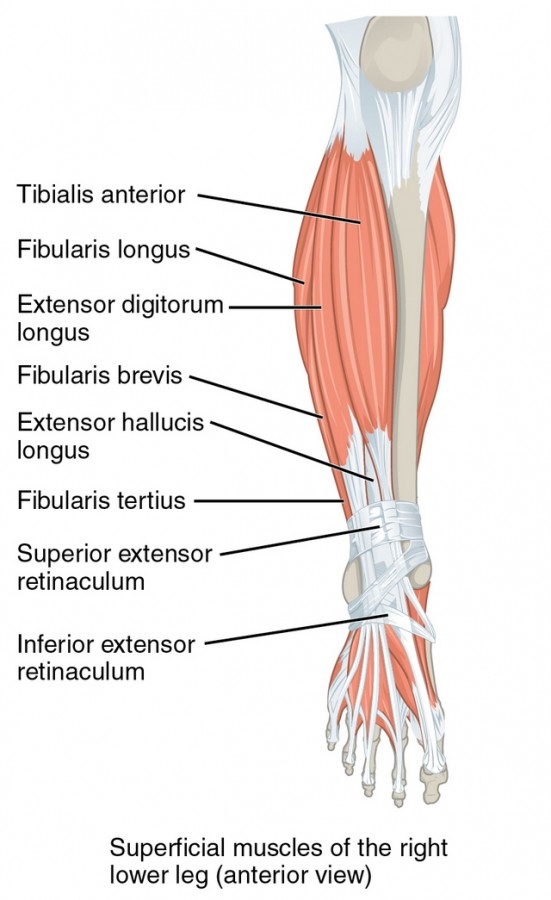
Tibialis Anterior Muscle
Tibialis anterior is a spindle-shaped muscle that originates from the lateral tibial condyle and, in parts, from the lateral surface of the tibia, the interosseous membrane of the leg and the intermuscular septum. It passes under the superior and inferior extensor retinaculum and is inserted into the first (medial) cuneiform bone and the first metatarsal bone. It is innervated by the deep fibular (peroneal) nerve (L5—S1).
The primary functions of the tibialis anterior muscle are dorsiflexion of the ankle joint and inversion (adduction/supination) of the foot. The tibialis anterior also stabilizes the ankle in the initiation of the stance phase.
In the case of weakness or damage of the deep fibular nerve, the ankle joint loses its dorsiflexion capabilities and the so-called foot drop occurs. It can be corrected with an ankle-foot orthosis (AFO) and treated with physiotherapeutic interventions.
Extensor Digitorum Longus Muscle
The extensor digitorum longus muscle has its origin from the lateral condyle of the tibia, the upper two-thirds of the fibula and the interosseous membrane of the leg. It inserts at the bases of the middle and distal phalanges of the 2nd to 5th toe. It is innervated by the deep fibular nerve (L4—S1) and expands the ankle joint dorsally.
The primary function of the extensor digitorium longus muscle is to dorsiflex the lateral four toes. It also synergizes the pronation in the lower ankle joint. Additionally, it dorsiflexes and everts (abducts/pronates) the foot.
Extensor Hallucis Longus Muscle
The extensor hallucis longus muscle originates from the middle half of the anterior surface of the fibula and the interosseous membrane and inserts on the base of the distal phalanx of the big toe (hallux). It is innervated by the deep fibular nerve (L5—S1) and extends the upper ankle joint.
The primary function of the extensor hallucis longus muscle is to dorsiflex the big toe. Additionally, it dorsiflexes and inverts (adducts) the foot.
Peroneus Tertius Muscle
The peroneus tertius muscle originates from the distal third of the fibula and the interosseous membrane. It inserts at the base of the 5th metatarsal bone. The peroneus tertius is inconsistent and is missing in about eight percent of the world population. Its insertion is sometimes added on the dorsal aponeurosis of the fifth toe or the fourth metatarsal. It is innervated by the deep fibular nerve (L4—S1).
The primary function of the peroneus tertius muscle is to dorsiflex and evert (abduct) the foot.
Plantar Flexors of the Upper Ankle Joint
The plantar flexors of the upper ankle joint are a group of muscles that includes the triceps surae, plantaris, tibialis posterior, flexor digitorum longus, flexor hallucis longus, peroneus longus and peroneus brevis.
With the exception of the peroneus muscles, which belong to a separate subgroup and are part of the lateral compartment of the lower leg, the plantar flexor muscles construct a part of the posterior compartment of the leg.
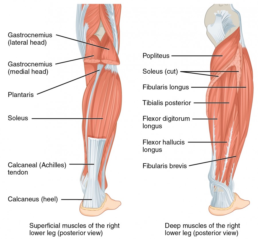
Triceps Surae Muscle Group
The triceps surae muscular unit consists of the gastrocnemius (medial and lateral heads) and the soleus muscle.
The medial and lateral heads of the gastrocnemius are originated proximally from the medial and lateral condyles of the femur, respectively. Both are inserted at the calcanealtuberosity via the calcaneal tendon. The gastrocnemius receives innervation through the tibial nerve (S1—S2) and is the strongest plantar flexor of the upper ankle joint.
The soleus is a small muscle that originates from the head and neck of the fibula and the tendinous arch of the tibia at the soleal line. It inserts at the calcaneal tuberosity via the calcaneal tendon and is innervated by the tibial nerve (S1—S2). It synergizes with the gastrocnemius to perform plantar flexion in the upper ankle joint.
In some people, the soleus muscle may additionally be supplied by the L5 segment. Next, to their primary function, the gastrocnemius and the soleus can supinate (invert/adduct) the lower ankle joint. The gastrocnemius muscle also flexes the knee.
Plantaris Muscle
The plantaris muscle originates from the lateral condyle of the femur (dorsal) and inserts at the calcaneal tuberosity. It is innervated by the tibial nerve (L4—S1) and acts as a plantar flexor of the upper ankle joint.
In some people, the plantaris muscle may additionally be supplied by the L4 segment. Besides its primary function, it is a flexor and internal rotator of the knee joint as well as a supinator (adductor) of the lower ankle joint.
Tibialis Posterior Muscle
The tibialis posterior muscle is originated from the interosseous membrane of the leg and the posterior borders of the tibia and fibula. It is inserted into the tuberosity of the navicular bone,the 1st—3rd cuneiform bones and the 2nd—4th metatarsal bones. It is innervated via the tibial nerve (L5—S1). In some people, tibialis posterior may be additionally supplied by the L4 segment.
The primary function of tibialis posterior is plantar flexion of the upper ankle joint. Additionally, it adducts (inverts) the lower ankle joint and performs tension of the transverse and longitudinal arch.
Flexor Digitorum Longus Muscle
Flexor digitorum longus originates from the middle third of the posterior surface of the tibiaand inserts on the plantar side of the bases of the lateral four (2nd—5th) distal phalanges. It receives its innervation from the tibial nerve (L5—S2).
The primary function of the flexor digitorum longus muscle is to flex the lateral four toes and plantar-flex the upper ankle joint. Additionally, it supports the foot’s longitudinal arch.
Flexor Hallucis Longus Muscle
Flexor hallucis longus finds its origin at the distal 2/3 of the posterior surface of the fibula and the interosseous membrane of the leg. It is inserted at the distal phalanx of the big toe (hallux)and is innervated by the tibial nerve (L5—S2).
As the name indicates, the primary function of this muscle is to flex the big toe and plantar-flex the ankle joint.
Peroneus (Fibularis) Longus Muscle
The long peroneus muscle originates from the lateral tibial condyle and the head and upper lateral side of the fibula and is inserted into the medial cuneiform bone and the base of the 1st metatarsal. It is innervated by the superficial peroneal (fibular) nerve (L5—S1), which is a branch of the common peroneal nerve.
The peroneus longus muscle serves as a plantar flexor of the upper ankle joint. It also everts (abducts/pronates) the foot and actively supports the transverse and longitudinal arch.
Peroneus (Fibularis) Brevis Muscle
The smaller peroneus muscle has its origin from the distal half of the lateral surface of the fibula and the anterior and posterior intermuscular spetum, while Its insertion is at the base of the 5th metatarsal. Its innervation is provided by the superficial fibular nerve (L5—S1).
The function of peroneus brevis is similar to that of peroneus longus. It plantar-flexes and everts the foot.
Pronators (or Abductors) of the Lower Ankle Joint
This group consists of specific muscles of the dorsiflexors and plantar flexors that have synergetic functions due to their functional anatomy. This group abducts (everts/pronates) the foot and includes the following muscles:
- Peroneus brevis: The peroneus brevis muscle is able to abduct the forefoot during contraction by simultaneously elevating the fifth metatarsal (outer margin of the foot). Due to the ligamentous connection, the cuboid bone moves along with the fifth metatarsal bone, as do the navicular bone and the calcaneus. The calcaneus also moves dorsally, causing a narrowing of the tarsal sinus.
- Peroneus longus: The long peroneus muscle pulls the forefoot laterally, while simultaneously lowering the medial edge of the foot. This is done through the connection between the medial cuneiform and the metatarsal bones.
- Extensor digitorum longus: It synergizes the movement of both peroneus muscles through its fibrous components.
Supinators (or Adductors) of the Lower Ankle Joint
This group also comprises of specific muscles of the dorsiflexor and plantar flexor groups that, in this case, have a synergistic effect when lifting the inner side of the foot. This group inverts (supinates/adducts) the foot and consists of the following muscles:
- Tibialis anterior: It pulls the foot into adduction by raising the inner edge of the foot. Its connection with the medial cuneiform and the first metatarsal also makes the entire forefoot follow this movement.
- Tibialis posterior: The tibialis posterior muscle is the most important contributor to the adduction movement. It pulls the navicular bone in medial direction, taking along the cuboid bone via ligamentous connections. Through its relation with the cuboid, the calcaneus also follows this medial movement and expands the tarsal sinus.
- Triceps surae: It synergizes the supination from the hindfoot. The hindfoot pulls the heel, and, therefore, the majority of its fibrous parts are shifted over the supinator-pronator axis. Biomechanically, its supination function is more pronounced.
Muscles of the Dorsum of the Foot
This group consists of only two muscles: the extensor digitorum brevis and the extensor hallucis brevis. Both of these muscles originate from the dorsal surface of the calcaneus.
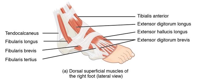
Extensor Digitorum Brevis Muscle
Extensor digitorum brevis originates from the dorsal surface of the calcaneus and inserts at the dorsal expansion of the medial four toes. It is innervated by the deep peroneal (fibular) nerve (L5—S1). This muscle functions as an extensor (dorsiflexor) of the medial four toes. It may also insert at the fifth toe and, therefore, be able to extend it.
Extensor Hallucis Brevis
The origin of extensor hallucis brevis is at the dorsal surface of the calcaneus, and it is inserted at the proximal phalanx and the dorsal expansion of the big toe (hallux). When actively innervated by the deep fibular nerve (L5—S1), it acts as an extensor (dorsiflexor) of the big toe.
The extensor hallucis brevis muscle is a part of extensor digitorum brevis and it can also synergize the extension of the other toes.
Muscles of the Sole of the Foot
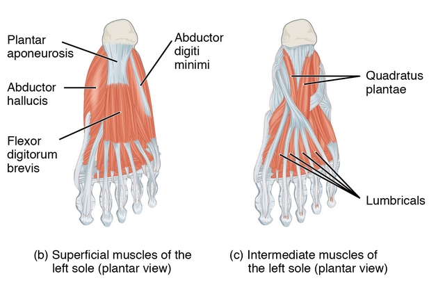
Flexor digitorum brevis, quadratus plantae, the lumbrical muscles and the dorsal and plantar interossei muscles form the large muscle group of the foot sole.
Flexor Digitorum Brevis Muscle
The flexor digitorum brevis muscle originates from the medial tubercle of the calcaneus and is inserted on the middle phalanges of the lateral four toes. It is innervated by the medial plantar nerve (L5—S1). It flexes the metatarsophalangeal and the proximal interphalangeal joints of the lateral four toes.
Quadratus Plantae Muscle
Quadratus plantae originates plantar from the calcaneus and inserts into the lateral border of the tendon of the flexor digitorum longus. It is innervated by the lateral plantar nerve (S1—S2) and acts as a plantar flexor of the toes.
Since the quadratus plantae muscle has no bony insertion, it synergizes the functions of the flexor digitorum longus muscle.
Lumbrical Muscles
The lumbrical muscles are four muscles that originate from the tendons of the flexor digitorum longus and insert medially into the proximal phalanges and the plantar aponeurosis. They are innervated by the medial and lateral plantar nerves (L5—S2). The medial plantar nerve innervates the first lumbrical, while lateral plantar nerve innervates the other three lumbricals.
The lumbricals plantar-flex the metatarsophalangeal joints and extend (dorsiflex) the interphalangeal joints.
Dorsal and Plantar Interosseous Muscles
The four dorsal interossei are bipennate muscles that originate from the adjacent metatarsal bones and insert into the proximal phalanx and the dorsal expansion of the second to fourth toe. When actively innervated by the lateral plantar nerve (S1—S2), they abduct the toes. They also flex the proximal and extend the distal phalanges.
The three plantar interossei are unipennate muscles that originate medially from the shafts of the 3rd – 5th metatarsals and are inserted at the base of the proximal phalanges and the dorsal expansion of the 3rd—5th toes. They are innervated by the lateral plantar nerve (S1—S2). The plantar interossei muscles adduct the toes and also flex the proximal and extend the distal phalanges.
The functions of the interossei muscles can be remembered by the mnemonic: PAD – DAB
PAD = Plantar interossei Adducts, while
DAB = Dorsal interossei Abducts.
Muscles of the Hallux
The ball of the big toe is formed by the abductor hallucis, the flexor hallucis brevis and the adductor hallucis.

Abductor Hallucis Muscle
The origin of abductor hallucis is at the medial process of the calcaneal tuberosity and the plantar aponeurosis. It inserts into the base of the proximal phalanx of the big toe. It is innervated by the medial plantar nerve (L5—S1). As the name indicates, it is an abductor of the big toe.
Flexor Hallucis Brevis Muscle
Flexor hallucis brevis originates from the undersurface of the cuboid and the three cuneiform bones and is inserted at the proximal phalanx of the big toe.
It is innervated by the medial plantar nerve (S5—S1) and flexes the big toe.
Adductor Hallucis Muscle
The adductor hallucis muscle has two origin heads: an oblique head and a transverse head. The oblique head mainly originates from the bases of the 2nd—4th metatarsals, while the transverse head originates from the capsule of the lateral four metatarsophalangeal joints. Both are inserted at the proximal phalanx of the big toe. It is innervated by the lateral plantar nerve(S1—S2) and adducts the big toe.
Muscles of the Little Toe
The ball of the little toe comprises of abductor digiti minimi, flexor digiti mimimi brevis and opponens digiti minimi.
Abductor Digiti Minimi Muscle
The abductor digiti minimi muscle originates from the medial and lateral processes of the calcaneal tuberosity and is inserted at the proximal phalanx of the little toe. When actively innervated by the lateral plantar nerve (S1—S2), it acts as an abductor of the small toe.
Flexor Digiti Minimi Brevis Muscle
Flexor digiti minimi brevis originates from the base of the 5th metatarsal and is inserted at the base of the proximal phalanx of the little toe. It is innervated by the lateral plantar nerve (S1—S2) and flexes the little toe.
Tabular Overview of the Muscles of the Lower Leg and Foot
Review Questions
Solutions can be found below the references.
1. Which of the following muscles is not a plantar flexor of the upper ankle joint?
- Long peroneus muscle
- Short peroneus muscle
- Flexor hallucis longus
- Plantaris
- Tibialis anterior
2. What muscles form the triceps surae?
- Gastrocnemius and plantaris
- Gastrocnemius and soleus
- Gastrocnemius and popliteus
- Gastrocnemius and long peroneus muscle
- Gastrocnemius and short peroneus muscle
3. Which nerve innervates the peroneus group?
- Sciatic nerve
- Tibial nerve
- Superficial fibular nerve
- Deep fibular nerve
- Femoral nerve


Comentários
Enviar um comentário