Nerve Tissue: Brain
In LECTURIO :) - Table of Contents

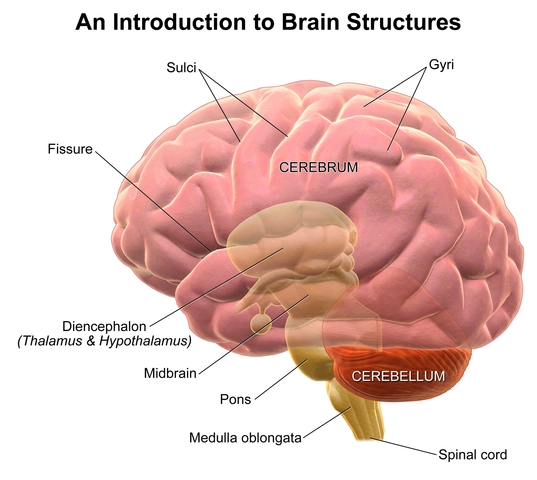
Introduction to the Brain
The brain is the central organ of the human nervous system along with the spinal cord. It is made up of:
- The cerebrum
- Diencephalon, consisting of thalamus, hypothalamus and pituitary gland
- The cerebellum
- The brainstem- consisting of midbrain, pons and medulla
The organ weighs about 1400 grams on average in an adult. It is housed and protected by the skull bones which include the:
- Parietal
- Frontal
- Occipital
- Ethmoid
- Sphenoid bone
- The bones of the face

At birth, the paired and unpaired bones of the skull are in close association to each other via the metopic, coronal, sagittal and lambdoid sutures. The sutures unite at the anterior and posterior fontanelle, all of which fuse normally before the age of two years.
Any form of fusion before the full development of the brain leads to microcephaly and limited growth and maturation of the brain. Thus, in adulthood, the bones are united at sutural areas such as the coronal suture that unites the frontal and parietal bones, sagittal suture that unites the two parietal bones and the lambdoid suture that unites the occipital and parietal bones.
The functions of the brain include:
- Control of the activities of the rest of the body
- Processing, integration, and coordination of information from the peripheral nervous system
- It is also involved in carrying out cognitive functions such as control of attention, preservation and memory, expression, and language, as well as visual functions
The Cerebrum
It is the largest part of the brain that is divided into the right and left cerebral hemisphere. The two hemispheres communicate with each other via the five commissures mainly through the corpus callosum.
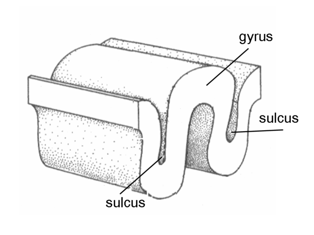
Image: “Gyrus Sulcus” by Albert Kok. License: Public Domain
The gross anatomy of the cerebrum involves anatomical and functional organization into a series of grooves and ridges, known as sulci and gyri, respectively. Each has been named differently as follows.
Important sulci include:
- Medial Longitudinal Fissure that separates the left and right cerebral hemispheres
- Lateral Sulcus that separates the parietal and temporal lobes
- Central Sulcus separating the parietal and frontal lobes
- Collateral Sulcus separating the fusiform gyrus and the hippocampal gyrus on the lower surface of the temporal lobes
- Calcarine Sulcus that separates the occipital lobes
Important gyri include:
- Anterior central gyrus
- Posterior frontal gyrus that is mainly involved in motor function
- Cingulate Gyrus which is located above the corpus callosum. It forms part of the limbic system
- Fusiform Gyrus which is in the temporal and occipital lobes consisting of lateral and medial parts. It is thought to play a role in facial and word recognition
- Inferior temporal gyrus that carries the auditory center
- Hippocampal Gyrus (Parahippocampal Gyrus) is a fold on the inner surface of the temporal lobe that borders the hippocampus. The hippocampal gyrus surrounds the hippocampus and plays an important role in memory
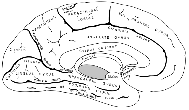
Image: “Medial surface of the left cerebral hemisphere.” by Henry Vandyke Carter – Henry Gray. License: Public Domain

Further division of the cerebrum gives the frontal, temporal, parietal, and occipital lobes of the brain. Microscopically, each hemisphere has an outer grey matter and an inner white matter. The white matter is further split into smaller divisions of the larger neocortex made up of 6 layers and the smaller allocortex made up of 4 layers.
Histologically, the cerebrum is arranged into layers comprising of Schwann cells, microglial cells, ependymal cells, and other types of neuronal cells.
The cerebrum is connected to the spinal cord by the brainstem which is made up of:
- The midbrain
- The pons
- The medulla oblongata
The cerebrum is further divided into telencephalon and diencephalon. The parts of telencephalon are:
- Cortex
- Subcortical fibers
- Basal ganglion
The parts of diencephalon are the:
- Hypothalamus
- Thalamus
- Pineal gland
- Pituitary gland
- Limbic structures (amygdala and hippocampus)
- The nuclei of the basal ganglia
The Cerebellum
This is the part of the brain that lies in the posterior fossa of the brain. It is connected to the brainstem via the superior cerebral peduncle that connects the cerebellum to the midbrain, the middle cerebella peduncle that connects the cerebellum to the pons and the inferior cerebella peduncle that connects the cerebellum to the medulla.
Grossly, it is divided into an anterior lobe, a posterior lobe and a flocculonodular lobe. The anterior and posterior lobes are connected via the cerebellar vermis.
As seen with the cerebrum, it also has an inner white matter known as the medulla and an outer grey matter known as the cortex.
The Meninges
The brain and spinal cord are surrounded by three layers of membranes known as meninges which are:
- An outer tough dura matter
- Delicate middle arachnoid layer
- An inner pia matter that is adherent to the brain and spinal cord
These meninges cover the brain and continue to cover the spinal cord to various levels beyond the foramen magnum where they exit.

The dura matter
It is the thick outermost layer in relation to the bones of the skull. It has two layers:
- An outer periosteal layer that is adherent to the periosteum and exits via various foramina to unite with the outer periosteum.
- An inner meningeal layer that is in close relation with the arachnoid layer.
It continues to cover the spinal cord beyond the foramen magna. At various levels, the dura projects into the cranial cavity to form various partitions. These projections include:
- The falx cerebri which partitions the right and left cerebral hemispheres. It runs from its anterior attachment to the crista galli to the posterior attachment on the tentorium cerebri; another partition of dura matter over the cerebellum.
- Tentorium cerebelli which lies over the cerebellum as a tent separating the posterior cerebrum from the cerebellum. It is attached anteriorly to the clinoid processes and posteriorly to the groves of the transverse sinuses on the occipital bone.
- Diaphragma Sella is a horizontally suspended dural matter fold that lies over the Sella turcica of the sphenoid bone. It has an opening for the infundibulum to pass through.
The two layers of dura matter enlarge to make up the dural venous sinuses which act as conduits for drainage of venous blood from the brain and its related structures. These sinuses include:
The arachnoid matter
This is the middle avascular membrane that is found in close relation to the dura. It has projections that run into the pia matter.
The pia matter
It is a delicate membrane that is found in close association with the brain and nerves exiting from the brain. It enters the various groves and fissures of the brain, unlike other membranes.
The folds of dura matter and the ability of the pia matter to enter various groves and fissures of the brain leads to the creation of various spaces within the membranous coverings of the brain.
The extradural space: Which is a potential space located between the dura and its tight adherence to the bones. Pathologically fluid, blood or other foreign bodies may accumulate in this space.
The subarachnoid space: It is a space dura matter and arachnoid matter, and between the pia matter and the overlying arachnoid matter. It normally contains cerebrospinal fluid. The various cavities enlarge to form cisterns that accumulate cerebrospinal fluid.
The cerebrum also contains the ventricular system which is made up of ventricular spaces that enlarge and contain free-flowing cerebrospinal fluid that is produced, accumulated, and finally absorbed in the ventricular system. The fluid suspends the brain and protects it against a sudden change in position and undue forces.
Normal flow of cerebrospinal fluid
CSF is produced within the cerebral ventricular system, mainly by the choroid plexus. The amount of CSF produced is 450 mL/day in the choroid plexuses. Each of the four ventricles contains choroid plexus tissue which is made up of villous folds lined by epithelium and a central core of highly vascularized connective tissue. Here, active secretion and diffusion produce CSF.
- From the two lateral ventricles, fluid drains through the foramen of Monro on each side into a single midline third ventricle.
- The third ventricle connects by the aqueduct of Sylvius to a midline fourth ventricle that has three exit openings, paired lateral foramina of Luschka, and a midline foramen of Magendie. These openings lead to a system of interconnecting and focally enlarged areas of subarachnoid spaces referred to as cisterns which allow for free flow of CSF.
- Fluid in the basal cisterns, tentorium, and subarachnoid space flows over the cerebral convexities to the sagittal sinus, from where absorption into the systemic circulation takes place.
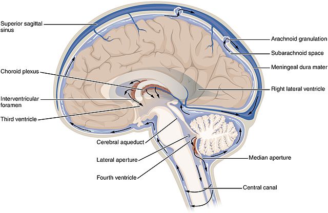
CSF absorption takes place mostly in the arachnoid villi into the venous channels of the sagittal sinus, but the fluid is also absorbed in the ependymal lining of the ventricular system and in the spinal subarachnoid space.
In the normal adult, the total volume of CSF is approximately 150 mLat a time, of which 25% is within the ventricular system. CSF is formed at the rate of 20 mL/h. The CSF production increases with a rise in intracranial pressure up to maximum levels.
Thus, there must be some absorption of the fluid to accommodate the volume of CSF formed each day.
The pathological state of hydrocephalus arises from:
- Increase in production of cerebrospinal fluid that may be caused by tumors of the ventricles such as choroid plexus papilloma and carcinomas.
- Reduction in the rate of absorption is seen in subarachnoid hemorrhage and meningitis that inhibit the brain to absorb CSF.
- Obstruction of the flow of cerebrospinal fluid, inhibit CSF to reach the sites where it is absorbed. Dilatation of the ventricles proximal to the obstruction is the manifestation of this pathway. Some of the common causes of obstruction include colloid cyst of the third ventricle obstructing at the foramen of monro, and tectal plate glioma obstructing and the cerebral aqueduct.
The Spinal Cord
The spinal cord is the anatomical connection between the brain and peripheral nervous system that begins at the foramen magnum as an extension of the brain to the lower margin of the L1 vertebral body in adults.
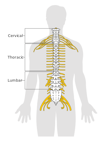
It is located within the bony vertebra which protects it and allows for division of the spinal cord into 31 spinal segmentswhich include:
- 8 cervical segments
- 12 thoracic segments
- 5 lumbar segments
- 5 sacral segments
- 1 coccygeal spinal segment
Each segment has a pair of:
- Anterior (motor) spinal nerve roots and
- Dorsal (sensory) spinal nerve roots that emerge from each side and combine to form the spinal nerve as it exits from the vertebral column through the neuroforamina.
The conical end of the spinal cord is known as the conus medullaris and followed by the spinal canal comprising of the lumbar, sacral, and coccygeal spinal nerves thereby forming the cauda equina.
A cross-section of the spinal cord reveals a central grey matter and a peripheral white mattercarrying various pathways of nervous tissue.
Sympathetic nervous system fibers exit the spinal cord between C7 and L1, while parasympathetic system pathways exit between S2 and S4. The sympathetic and parasympathetic fibers supply the bladder, while the motor and sensory fibers below the L2 innervate the perineal region, the lower limbs and part of the pelvis.


#/media/File:Gyrus_sulcus.png)
Keep it Sir and I am waiting for your next post on your site blog.
ResponderEliminarhttps://blog.mindvalley.com/lobes-of-the-brain/
Great writing it is such a cool and nice blog, thanks for sharing your post. I like your post very much. Thanks for your post.
ResponderEliminarhttps://blog.mindvalley.com/function-of-occipital-lobe/