Chambers and Great Vessels of the Heart
Table of Contents
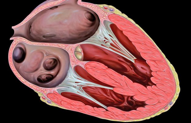
Image: “Heart transesophageal echocardiography (TEE) view.” by atrick J. Lynch, medical illustrator – Patrick J. Lynch, medical illustrator. License: CC BY 2.5
Chambers of the Heart
Atria
The atria are located on the anterior aspect of the heart and receive blood. The superior and inferior vena cava transport oxygen-poor blood from the systemic circulation to the right atrium, which then pumps it into the right ventricle.
The left and right pulmonary veins transport oxygen-rich blood from the pulmonary circulationto the left atrium, which then sends it across the mitral valve to the left ventricle.
The atria receive blood passively, i.e. the blood does not traverse through any valves. During atrial diastole (relaxation), the atria dilate and are filled with blood, and during atrial systole (or contraction) they pump the blood into the ventricles. The atria promote continuous flow of venous blood as there are:
- No atrial valves
- Contractions during atrial systole are incomplete, thereby allowing venous blood to flow uninterruptedly
- Atrial contractions are mild, there is no back pressure to prevent venous blood flow
- Atrial diastole begins prior to the start of the ventricular systole.
Ventricles
The two ventricles of the heart are situated on its posterior aspect, below their corresponding atrium. The left atrium pumps oxygen-rich blood to the left ventricle during diastole. The left ventricle then pumps the blood into the aorta, which then transports it via its branches to the systemic circulation.
The right atrium pumps oxygen-poor blood into the right ventricle, which then transports it via the pulmonary vein to the pulmonary circulation for oxygenation.
The mitral valve separates the left atrium and ventricle while the tricuspid valve separates the right atrium and right ventricle. The valves are attached to the chordate tendinae, which are elastic tendons that open the valve during atrial systole/ventricular diastole but close the valve during ventricular systole preventing regurgitation of blood.
The ventricles have thicker, more muscular walls compared to the atria and the left ventricle has thicker walls compared to the right ventricle. There are semilunar valves at the opening to the aorta and the pulmonary vein. These valves close during ventricular diastole and separate the left ventricle from the aorta and the right ventricle from the pulmonary vein, thus preventing regurgitation of blood.
The volume of blood pumped by the left ventricle to the systemic circulation is larger compared to the volume of blood pumped by the right ventricle to the pulmonary circulation.
Great Vessels of the Heart
The great vessels of the heart are all located in the mediastinum. They help to transport blood between the heart and the rest of the body. There are five great vessels, which enter and leave the heart. These are the superior and inferior vena cava, which bring oxygen-poor blood to the heart, the aorta, which carries oxygen-rich blood from the heart, the pulmonary artery and thepulmonary vein.
Superior vena cava
It is a large vein, which brings oxygen-poor blood from the upper arms, and head–neck region to the right atrium. The merger of the brachiocephalic veins posterior to the meeting point of the first costal cartilage and the sternum leads to the formation of the superior vena cava.
The two brachiocephalic veins themselves are formed when the internal jugular and the subclavian veins merge. The thoracic duct drains into the left subclavian vein on the left side of the body. So the superior vena cava also recirculates lymph.
Inferior vena cava
This is a large vein, which brings oxygen-poor blood from the lower limbs, pelvis and the torso to the right atrium. It is formed when the right and the left common iliac veins merge at the L5 vertebral level.
It ascends along with the abdominal aorta, and then passes through the diaphragmatic cavalhiatus at the T8 vertebral level. The right gonadal, right suprarenal vein, the left renal vein,and all the lumbar, hepatic and phrenic veins drain into the inferior vena cava.
Pulmonary arteries
The pulmonary arteries are responsible for transporting oxygen-poor blood from the right ventricle for oxygenation to the pulmonary alveolar capillaries. The right ventricle transports blood across the semilunar valve into the solitary pulmonary trunk, which subsequently separates into the right and left pulmonary arteries at the T5-T6 level.
The right atrium lies posterolateral to the pulmonary trunk, which overlaps the aortic root as it rises superiorly and posteriorly. The left pulmonary artery is shorter and narrower compared to the right pulmonary artery.
Pulmonary veins
The pulmonary veins transport oxygen-rich blood to the left atrium from the pulmonary circulation. The function is carried out by four veins – the superior pulmonary veins carry blood from the superior pulmonary lobes while the inferior pulmonary veins carry blood from the inferior pulmonary lobes.
The superior vena cava and the right atrium lie anterior to the right pulmonary veins while thedescending thoracic aorta lies posterior to the left pulmonary veins.
Aorta
The aorta carries oxygen-rich blood to the entire body from the left ventricle. The left ventricle transports blood across the semilunar aortic valve into the ascending part of the aorta, which then continues as the aortic arch, then as the thoracic aorta and finally terminates as the abdominal aorta at the L4 vertebral level.
Ascending aorta
The ascending aorta is 2 inches in length and travels in the pericardial sheath along with the pulmonary trunk. It has the right and left aortic dilatations or sinuses near the aortic valve. These give rise to the right and left coronary arteries.
Aortic arch
The second intercostal joint is the landmark where the ascending aorta continues as the aortic arch, which then travels posterosuperiorly and then inferiorly. Subsequently, it descends in the posterior mediastinum on the left side of the midline. At the T4 vertebral level, the aortic arch becomes the thoracic aorta.
The aortic arch gives off the following branches: the right brachiocephalic trunk , the left common carotid artery and the left subclavian artery.
Thoracic aorta
This begins on the left side of the midline as a continuation of the aortic arch and runs from T4 to T12 vertebra. As it nears the aortic hiatus in the diaphragm, it lies near the midline. It gives off the following branches: bronchial, mediastinal, esophageal, pericardial, superior phrenic, intercostal and subcostal arteries.
Descending abdominal aorta
The thoracic aorta continues as the descending aorta at the aortic hiatus in the diaphragm at the T12 vertebral level and ends opposite the L4 vertebra. It terminates by branching into the two common iliac arteries. Its other branches are: inferior phrenic arteries, celiac arteries, superior mesenteric artery, middle suprarenal arteries, renal arteries, gonadal arteries, inferior mesenteric artery, median sacral artery, lumbar arteries.
Clinical Relevance
Aortic aneurysm
Aortic aneurysm is a dilatation of the arterial wall more than 50% of its diameter. This can occur in the aorta due to several causes. Thoracic aortic aneurysms are usually associated with Marfan’s syndrome while abdominal aortic aneurysms are associated with atherosclerosis and smoking. If untreated, the aneurysms can rupture, leading to death.
Dissection of the aorta
Dissection of the aorta refers to separation of the layers of the intima and media of the aortic wall. This causes abnormal flow of blood through two parallel channels with thinning of the vessel walls. It can subsequently result in the formation of an aneurysm.


Comentários
Enviar um comentário