The Ventricular System of the Brain
Table of Contents
- The Cerebral Ventricles
- Topography of the Ventricular System – The Inner Cerebrospinal Fluid Spaces
- Functions of the Ventricular System
- Circulation of Cerebrospinal Fluid Inside the two Cerebrospinal Fluid Spaces
- Circumventricular Organs
- Tissue Barriers inside the Brain
- Diseases of the Ventricular System
- Review Questions
- References
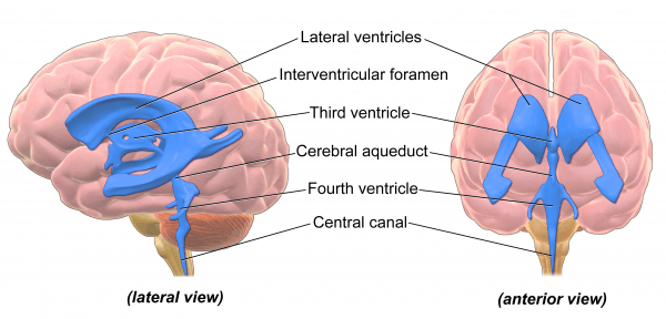
The Cerebral Ventricles
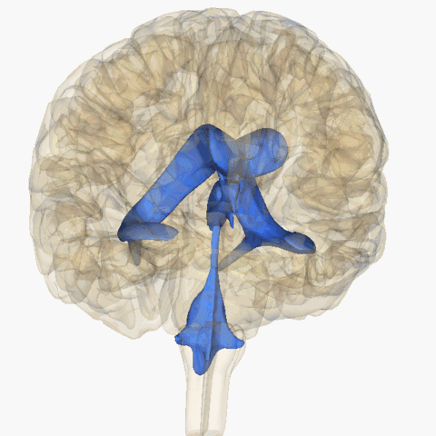
Image: “Rotating animation of ventricular system consisting of four ventricles.” by Life Science Databases (LSDB). License: CC BY-SA 2.5
The third ventricle (ventriculus tertius) is connected to the fourth ventricle through the aquaeductus mesencephali cerebri so that all ventricles are connected to each other. All ventricles together form the inner cerebrospinal fluid spaceand are connected to the outer cerebrospinal fluid space, the subarachnoid space (spatium subarachnoidem), through the fourth ventricle via three apertures.
A total of 150ml of cerebrospinal fluid circulates through the two cerebrospinal fluid spaces, of which there are approximately 30ml of cerebrospinal fluid in the inner and approximately 120ml of cerebrospinal fluid in the outer cerebrospinal fluid space.
The cerebrospinal fluid is produced primarily by the plexus choroideus, which can be found in each of the four ventricles but it is also partly produced by the specialized epithelium of the ventricles, the ependyma. The entire inner cerebrospinal fluid space is lined with ependyma.
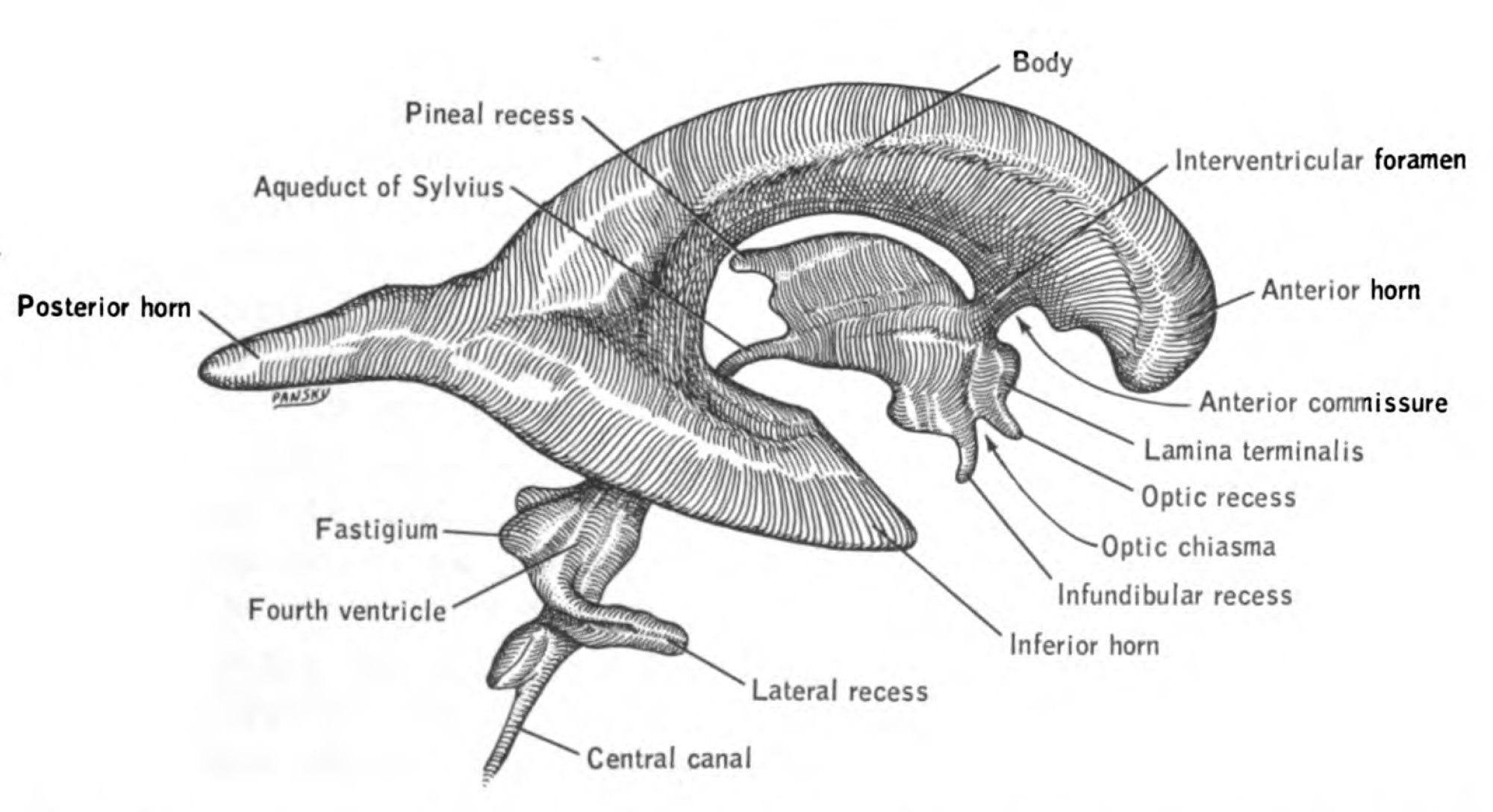
Image: “Anatomical illustration of the human nervous system from the 1960 book – A functional approach to neuroanatomy” by House, Earl Lawrence. Pansky, Ben. License: Public Domain
Topography of the Ventricular System – The Inner Cerebrospinal Fluid Spaces
Topography of the Ventriculi Laterales
The two lateral ventricles form the largest cavities of the ventricular system and can be subdivided further. It is differentiated between the anterior horn (cornu frontale, anterius) the inferior horn (cornu temporale, inferius) and the posterior horn (cornu occipitale, posterius). The individual parts are assigned specific areas of the brain, depending on their topographic location.
The anterior horn is in close connection to the frontal lobe, the inferior lobe is assigned to the temporal lobe and the posterior horn to the occipital lobe. Important structures that border the lateral ventricles are the nucleus caudatus, which is located at the lateral front wall of the anterior horn, the thalamus, which is located at the lateral back wall of the anterior horn as well as the putamen, which does not border the lateral ventricle directly but is located laterally to it. The two lateral ventricles are each connected to the unpaired third ventricle through the foramen interventriculare. The foramen interventriculare is also referred to as foramen of Monro.
Topography of the Ventriculus Tertius
The third ventricle is associated with the interbrain (diencephalon = epithalamus, thalamus, and hypothalamus), which borders at its lateral wall. Especially to be emphasized in this context are the two thalami, which can touch each other in the area of the adhaesio interthalamica. However, this is not a functional connection meaning that there are no commissure fibers between the two thalami. The aquaeductus mesencephali cerebri, which connects the third and the fourth ventricle, is attributed to the mesencephalon.
Topography of the Ventriculus Quartus
The fourth ventricle is associated with the hindbrain (rhombencephalon = pons, cerebellum, medulla oblongata). The ventriculus quartus functions as a connection to the outer cerebrospinal fluid space through its apertures. Here, one differentiates between pairedaperturae laterals (Luschkae) and unpaired aperture mediana (Magendi). Parts of the plexus choroideus even reach from the aperturae laterales into the subarachnoid space. These parts are also referred to as the flower basket of Bochdalek or Bochdalek’s flower basket. The fourth ventricle is connected to the spinal canal via the canalis centralis.
Functions of the Ventricular System
A major function of the ventricular system is the production of cerebrospinal fluid, which represents the ultra-filtration of blood that is produced by the plexus choroidei of the four ventricles. The cerebrospinal fluid protects the brain against mechanical impacts by reducing the effective weight of the brain considerably. This is made possible because the brain swims in cerebrospinal fluid (buoyancy principle), so to speak. Furthermore, the cerebrospinal fluid serves to supply the brain with nutrients and disposes of metabolites.
Structure of the Plexus Choroideus
The plexus choroideus is the main production site of cerebrospinal fluid and can be found in all four ventricles. Inside the two lateral ventricles, the plexus choroideus originates from the base of the pars centralis, as well as from the roof of the inferior horn. Inside the third ventricle, the plexus choroideus is located in the roof and inside the fourth ventricle it is found on the back wall, as well as below the cerebellum.

Image: “Choroid Plexus Histology” by Marvin_101. License: CC BY-SA 3.0
The plexus choroideus is formed by protrusions of the ventricle wall and by the ingrowth of capillary loops into the ependyma of the ventricle. Consequently, the plexus choroideus is a fixed component of the ventricle wall and can only be separated mechanically. If the plexus choroideus is removed with forceps, tear-off lines, also referred to as taenien, will appear on the ventricle wall. The surface of the plexus choroideus shows numerous folds and an apical brush border, both of which serve to enlarge the surface.
The epithelium of the plexus itself is a single-layer cuboidal epithelium. The layer of connective tissue that forms the plexus choroideus contains a lot of vessels and is also referred to as tela choroidea. Approximately 500ml of cerebrospinal fluid are produced per day.
Circulation of Cerebrospinal Fluid Inside the two Cerebrospinal Fluid Spaces
The liquor cerebrospinalis is produced in all four ventricles inside the inner cerebrospinal fluid space, and transported via cilia carrying epithelium (ependyma). Via the apertura mediana and the paired aperturae laterales of the fourth ventricle, the cerebrospinal fluid passes from the inner to the outer cerebrospinal fluid space meaning from the ventricular system to the subarachnoid space.
The subarachnoid space shows numerous extensions that are also referred to as cisterns in which the cerebrospinal fluid circulates. The most important cisterns in this context are the cisterna cerebellomedullaris (=cisterna magna), the cisterna ambiens, the cisterna interpeduncularis, and the cisterna chiasmatica.
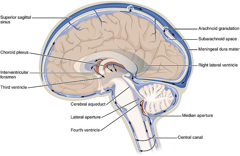
Clinically important are especially those cisterns that can be used to extract cerebrospinal fluid via puncture. During a lumbar puncture, the cisterna lumbalis is punctured; during a suboccipital puncture the cerebrospinal fluid is taken from the cisterna cerebellomedullaris. An indication for a lumbar puncture is given, for example, in cases of suspected meningitis.
There are two ways for the drainage of the subarachnoid space. On one hand, the cerebrospinal fluid flows across the granulationes arachnoideae (=Pacchioni granulations) into the venous plexus or the lymphatic system, on the other hand it is drained via the exits of the spinal nerves. The main drainage pathway, however, is via the granulationes arachnoideae. The entire cerebrospinal fluid (approximately 150ml) is exchanged two to four times a day in this cycle.
Summary of the circulation of the cerebrospinal fluid: ventricle (inner cerebrospinal fluid space) – via apertures of the fourth ventricle – subarachnoid space (outer cerebrospinal fluid space) – granulationes arachnoideae/exits of spinal nerves – venous vessel system.
Circumventricular Organs
The aforementioned plexus choroideus belongs to the group of circumventricular organs or ependymal organs, respectively. What makes this group so special is the fact that these regions usually do not contain a blood-brain-barrier.
An exception in this context is the subcommissural organ (organum subcommissurale), which is also part of the circumventricular organs even though the blood-brain-barrier exists here. Examples of circumventricular organs are:
- Posterior pituitary (neurohypophysis)
- Corpus pineale
- Organum vasculosum laminae terminales
- Organum subfornicale
- Area postrema
Tissue Barriers inside the Brain
The tissue barriers of the brain protect the brain from harmful substances in the blood. This also includes substances given via iatrogenic administration like drugs.
The blood-brain-barrier
The blood-brain-barrier mainly consists of “tight junctions” that are built by the endothelial cells of the capillaries. Transporters along the blood-brain-barrier allow the transport of essential substances for the brain across the barrier. Glucose, for example, can be transported to the brain via these transporters.
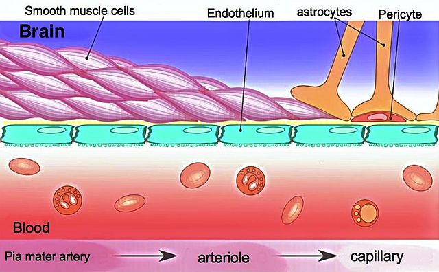
There is no blood-brain-barrier in circumventricular organs (see above) because the capillary endothelium is fenestrated in that area. To still have a barrier between brain tissue and cerebrospinal fluid in those areas, a blood-liquor-barrier is formed (see below).
The blood-liquor-barrier
The specialized ependymal cells of the plexus choroideus are connected to each other via “tight junctions” so that a barrier between blood and cerebrospinal fluid is formed in that area, but not between cerebrospinal fluid and CNS. This barrier can only be penetrated by a few substances like water, oxygen or carbon dioxide.
Diseases of the Ventricular System
Hydrocephalus
A pathologic enlargement of the cerebrospinal fluid spaces is called hydrocephalus (“Water head”). Depending on whether the inner or the outer cerebrospinal fluid space is enlarged, one refers to a hydrocephalus internus or externus, respectively. Hydrocephalus internus are caused especially by a failure to drain cerebrospinal fluid.

Particularly vulnerable for drainage failures are physiological constrictions within the ventricular system like the aquaeductus mesencephali or the apertures of the fourth ventricle. Possible reasons for a constriction are tumors, bleedings but also inflammations. Based on the pathogenesis, this type of hydrocephalus is also referred to as hydrocephalus occlusus.
An enlargement of the subarachnoid space (hydrocephalus externus), occurs especially during the physiological ageing process and the subsequent reduction of the cerebral parenchyma(atrophy). What is special about this type of hydrocephalus, which is also referred to as hydrocephalus e vacuo, is the fact that there is no increased cerebral pressure.
Adhesion of arachnoid villi and the resulting cerebrospinal fluid re-absorption failure can lead to an enlargement of the inner, as well as the outer, cerebrospinal fluid spaces. This type of hydrocephalus is also referred to as hydrocephalus malresorptivus or aresorptivus, respectively, based on its pathogenesis.
If an increased production of cerebrospinal fluid is the cause of a hydrocephalus, it is also called hydrocephalus hypersecretorius. This kind of over-production of cerebrospinal fluid can be a result of irritation of the plexus epithelium, for instance, as a result of an inflammation. Clinically, symptoms like headache, vomiting or nausea can be observed due to the increased cerebral pressure.
Opposite that is the normal pressure hydrocephalus where no increased cerebral pressure is measured. This type of hydrocephalus results in the typical clinical triad of gait disorder, urinary incontinence, and dementia, which are also called Hakim’s triad.
Therapy of a Hydrocephaly
Therapy focuses on the removal of possible reversible causes, i.e. an inflammation, as well as on the removal of eventual constrictions. Another possible therapy is shunting where cerebrospinal fluid is artificially drained from the ventricular system. This drainage can be done inside the peritoneal space (ventriculoperitoneal shunt), for instance, or inside the right atrium (ventriculoatrial shunt). An indication for shunting is given in cases of pediatric hydrocephalus or normal pressure hydrocephalus.
Review Questions
The answers are below the references.
1. Which statement concerning the ventricular system is true?
- One differentiates between five ventricles.
- The lateral ventricle is paired.
- The third and fourth ventricles are connected via the interventricular foramen.
- The connection between lateral ventricles and the third ventricle is called Aquaeductus.
- The ventricular system represents the outer cerebrospinal fluid space.
2. Which statement concerning the topography of the ventricles is not true?
- The anterior horn of the lateral ventricles is assigned to the frontal lobe.
- The Aquaeductus is attributed to the mesencephalon.
- The third ventricle is attributed to the diencephalon.
- The fourth ventricle is located inside the rhombencephalon.
- The fourth ventricle has no connection to the spinal cord canal.
3. Which statement concerning the apertures of the fourth ventricle is not true?
- There are three different apertures.
- The apertura mediana is paired.
- The apertura mediana is also referred to as foramen of Magendi.
- Cerebrospinal fluid is drained via the apertures into the subarachnoid space.
- The aperturae laterales are also referred to as foramen of Luschka.
4. Which statement concerning the plexus choroideus is true?
- It is located only inside the two lateral ventricles.
- The plexus epithelium is stratified.
- The plexus epithelium is only loosely connected to the ventricle wall.
- It is the major production site of cerebrospinal fluid.
- It consists of elastic fibers.


Comentários
Enviar um comentário