The Hepatic Portal System
Table of Contents
A Second Capillary Bed as a Safety System
When the blood passes through the gastrointestinal tract, it absorbs numerous substances from the food. This may include vital nutrients (except fats that enter the blood via the lymphatic system), but also dangerous and harmful foreign particles and pathogens.
One of the important functions of the liver is the detoxification of the body. The blood carries the many substances from the gastrointestinal tract directly into the liver and where they undergo biotransformation.
This way, harmful substances can be rapidly neutralized. In pharmacology, the presence of a first pass effect must be taken into account when administering oral medications, and drug dosage must be adjusted to compensate for this effect. If it is too low, the majority of the drug is metabolized in the liver and is no longer effective.
The liver must filter the blood from the spleen as well, since it is well known that red blood cells are degraded here. Their constituents accumulate in the blood and must be completely disassembled and recycled in the liver.
The Portal Vein System of the Liver
In addition to the stomach, small and large intestine, the blood from the gall bladder, pancreas and spleen also flows to the liver via the portal vein (technically called the hepatic portal vein). The portal vein drains blood delivered to the above organs via the three large, unpaired branches of the aorta: The Celiac Trunk, the Superior Mesenteric Artery and the Inferior Mesenteric Artery.
The hepatic portal vein is formed by the joining of the splenic vein and the superior mesenteric vein. The inferior mesenteric vein drains into the splenic vein. The majority of the blood from the abdomen runs through these three large veins. The following branches drain directly into the portal vein:
- Cystic vein
- Right and left gastric veins
- Pancreatico-duodenal superior posterior vein
- Pre-pyloric vein
- Para-umbilical vein
Since the blood is only partially saturated with oxygen, the liver needs additional oxygenated blood to supply its needs. The hepatic artery from the celiac trunk assumes this task. The portal vein is therefore the vas public of the liver, a vessel carrying blood that contains chemicals to be transformed for the good of the whole body. The portal vein and the hepatic artery, together with the common bile duct, the main duct carrying bile, form the porta hepatis.
After the blood has been processed, it finds its way into the inferior vena cava via the hepatic veins. Their vascular tree collects blood from the centrolobular veins of the hepatic sinusoids after the vessels of the porta hepatis have delivered the blood to their intrahepatic ramifications (see under liver).
Portal-systemic Anastomoses
When the liver is damaged however, for example in cirrhosis from chronic alcohol abuse, the vascular bed in the hepatic parenchyma is reduced. Therefore at a constant flow rate the same volume of blood must be pushed out of the portal vein via fewer vessels. The pressure in the portal vein increases and portal hypertension develops.
This may be so great that the blood reverses its direction of flow. It does not flow directly into the liver anymore, but rather away from the liver. It seeks other ways back to the heart. These are called the portal-systemic anastomoses:
1. Esophageal Anastomoses:
Portal vein → Gastric vv → Esophageal vv → azygos/hemiazygos → superior vena cava
2. Paraumbilical Anastomoses:
a) Portal vein → umbilical veins → paraumbilical vv → superior epigastric v. → internal thoracic v. → subclavian vein → superior vena cava
b) Portal vein → umbilical veins → paraumbilical vv → inferior epigastric → external iliac vein → inferior vena cava
c) Portal vein → umbilical veins → paraumbilical vv → superficial abdominal veins: thoracoepigastric v., lateral thoracic v., superficial epigastric vein
3. Colic Anastomoses:
Portal vein → superior and inferior mesenteric vein → Colic vv. → ascending lumbar vv. → directly into the inferior vena cava or via V.azygos/hemiazygos → superior vena cava
4. Rectal Anastomoses:
Portal vein → inferior mesenteric vein → superior rectal (hemorrhoidal) vv.→ middle/inferior rectal (hemorrhoidal) vv. → internal iliac vein → inferior vena cava
Usually the aforementioned collateral circulation has to do with small vessels, which are not suitable for larger amounts of blood. Due to the increased pressure they expand and can become visible.
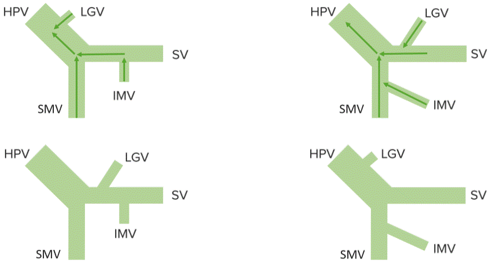
“Hepatic portal vein. Formation variation” Image by Lecturio
The anastomosis through the veins of the esophagus forms esophageal varices. These thin veins may stretch to the point that they can burst, causing an upper intestinal hemorrhage. Chronic bleeding causes anemia, acute situations can be life-threatening (see 1).
A phenomenon on the skin of the abdomen in which protruding veins around the umbilicus develop is called caput medusae. They arise through the bypass circuit over the re-opened umbilical veins (see 2).
The paraumbilical vein passes along the round ligament of the Livre to join with superficial epigastric veins which drain into the external iliac. If these become dilated the surface of the skin is elevated these varicose veins radiate from the umbilicus as caput-medusae (see 3).
In case of the colic vein, small branches from the colic veins unite with retroperitoneal veins that drain into the inferior vena cava (see 3).
The overfilling of the rectal veins causes hemorrhoids. These can also be extended to such an extent that they burst (see 4).
To decompress the portal system in cases of severe hemorrhage but a relatively preserved liver function, shunting procedures have emerged. The most common are:


Comentários
Enviar um comentário