Joints of the Lower Limb
Table of Contents
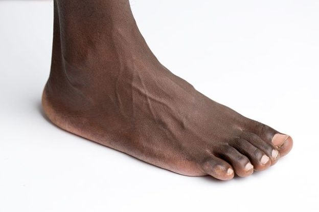
Image : “Foot on white background” by
Genusfotografen. License: CC BY-SA 4.0
Sacroiliac Joint
The two sacroiliac joints are formed between the articulating surfaces of the sacrum and the ilium. It is a synovial plane joint that transmits the weight of the body to the lower limbs thus it is a strong joint with very few movements between it.Strong ligaments that offer support to the joint include:
- Anterior sacroiliac ligament.
- Posterior sacroiliac ligament
- Sacro tuberous ligament
- Sacrospinous ligament.
Very little movement is permitted at these two joints.
Hip Joint
The hip joint is a synovial ball and socket joint formed between the head of the femur and the acetabulum. The acetabular labrum is a strong fibrocartilaginous ring along the edge of the acetabulum, which provides depth to the socket and helps to hold the femoral head within the socket.
The hip joint is designed as a weight bearing joint; therefore, strong ligaments stabilize it including:
- The ischiofemoral ligament.
- The iliofemoral ligament
- The pubofemoral ligament
- The ligament of the head of the femur.
Several bursae around the hip joint prevent friction between the joint and the strong musclessupporting it. These include the trochanteric bursa, the ischial bursa and the iliopectineal bursa. The different movements occurring at the hip joint include flexion, extension, abduction, adduction, medial and lateral rotation.
Knee Joint
The knee joint is a modified, compound hinge joint and consists of two articulations: one between the femur and the tibia and the second between the femur and the patella.
The joint is stabilized by strong muscles and ligaments which include the quadriceps and the hamstring muscles, the anterior and posterior cruciate ligaments, the medial collateral and the lateral collateral ligaments and finally the patellar ligament.
Cushioning the articular surfaces of the joint are the medial and lateral menisci, which are semi-lunar shaped fibrocartilages. Several bursae prevent friction between the structures supporting the knee joint: the suprapatellar bursa and the gastrocnemius bursa.
Movements occurring at the knee joint include flexion, extension, and a small degree of medial and lateral rotation. It is an important joint that helps to transmit the weight of the body to the foot. It is important for our daily activities such as walking, running, and sitting.
Tibiofibular Joints
There are two joints between the tibia and the fibula:
- The proximal tibiofibular joint/ superior tibiofibular joint is an arthrodial joint between the head of the fibula and the lateral condyle of the fibular. The articular facets are covered by cartilage connected by articular capsules.The proximal joint allows a gliding movement
- The distal fibrous syndesmosis. An interosseous membrane also connects the shafts of the tibia and fibula. The distal joint does not permit any movement.
Foot Joints
Ankle joint
Also called the talocrural joint, this is a true hinge joint formed between the articular surfaces of the distal tibia, distal fibula, and the talus. This functionally a hinge joint allowing plantar flexion, dorsiflexion and a small degree of abduction, adduction, and rotation.
Subtalar joint (talo-calcanean joint)
It is located distal to the ankle joint and is a synovial joint formed by the talus and the calcaneus bone. These two bones articulate twice: anteriorly and posteriorly when the concave area of the talus meets the convex surface of the calcaneus.
There are three articular facets between the talus and the calcaneus called the anterior, middle and the posterior facets. The middle facet is formed by the sustentaculum tali. The tarsal canal separates the posterior facet from the other facets.
The joint capsule is attached to the margins of the articular surface of both the bones. The ligaments which stabilize this joint are the medial, lateral and interosseous talocalcaneal ligaments.
10% of the ankle dorsiflexion is due to the subtalar joint. The joint mainly allows inversion andeversion of the foot.
Transverse tarsal joint (Chopart’s or mid-tarsal joint)
This joint consists of two joints: the talocalcaneonavicular and the calcaneocuboid. More extensive movements take place at this joint compared to other tarsal joints. It consists of a rotator movement whereby the foot may be slightly extended or flexed while the sole is being everted or inverted.
Talocalcaneonavicular joint
This is a synovial, modified ball and socket joint formed by the talus (representing the ball), the calcaneus and the navicular bones (forming the joint socket). The plantar calcaneonavicular ligament stabilizes this joint.
Calcaneocuboid joint
This joint is formed between the anterior aspect of the calcaneus and the posterior surface of the cuboid bone. It is a synovial, saddle type of joint stabilized by the bifurcated ligamentsuperiorly, the long plantar ligament inferiorly and the short plantar ligament deep to the long plantar ligament. The joint capsule is attached around the margins of the articular surface. This joint permits 300 of inversion and 200 of eversion.
Cuneonavicular joint
This joint formed by the navicular and the three cuneiform bones is a synovial joint which permits gliding movement. The dorsal and plantar cuneonavicular ligaments stabilize the joint.
Cuboideonavicular joint
This is a joint between the cuboid and navicular bones and is supported by the plantar, dorsal and interosseous ligaments. It is a fibrous joint.
Tarsometatarsal joint
These are synovial joints formed between the three cuneiform, cuboid and the bases of the (1st to the 5th) metatarsal bones. The dorsal, plantar and interosseous ligaments stabilize these joints.
Intermetatarsal joint
These are synovial joints between the bases of the metatarsal bones. The plantar, dorsal, and interosseous ligaments stabilize them.The base of the first metatarsal is not connected to the second via any ligaments.
Metatarsophalangeal joint (MTP)
These are joints between the metatarsal heads and the proximal phalange bases of the toes. The joints are synovial and are supported by the collateral and plantar ligaments. Due to these joints, we are able to flex and extend our toes, as well as adduct and abduct them to keep them apart or bring them closer.
Interphalangeal joint (IP) of the big toe
These synovial joints connect the phalanges of the big toe and are supported by the plantar and collateral ligaments. It allows flexion and extension of the big toe.
Proximal interphalangeal joint (PIP)
This joint is formed between the proximal phalanges and the middle phalanges of the four lateral toes.
Distal phalangeal joint (DIP)
These are joints between the middle and distal phalanges of the four lateral toes.
Plantar Ligaments
There are several ligaments which maintain the functional integrity of the sole of the foot. These are called the plantar ligaments.
- The long plantar ligament connects the calcaneus and the cuboid bone. It is the longest ligament of the tarsus and converts the groove on the cuboid into a canal for the fibularis longus tendon. The short plantar ligament is deep to the long plantar ligament.
- The plantar calcaneocuboid ligament lies deep to the long plantar ligament and is a short, wide band with great strength.
- The plantar calcaneonavicular ligament connects the calcaneus to the navicular bone.
- The plantar cuneonavicular ligament is between the navicular bone and the cuneiform bones.
- The Plantar Intercuneiform ligaments connect the cuneiform bones.
- The Plantar cuboideonavicular ligament connects the cuboid and navicular bone.
- The Plantar cuneocuboid ligament lies between the cuboid and the cuneiform bones.
Arches of the Foot
There are three arches in the foot: the medial longitudinal arch, the lateral longitudinal arch,and the transverse arch. The arches of the foot, along with the bones, joints, muscles and ligaments, play an important role in helping the foot absorb shock during walking, jumping and running.
The transverse arch is formed by the cuboid, the three wedge-shaped cuneiform bones and the bases of the first to the fourth metatarsal bones. It creates the medial to lateral mid-foot curvature which enables distribution of body weight within the foot from side to side and navigates uneven ground. It also provides elasticity to the foot enabling propulsion when walking.
The medial and lateral longitudinal arches are formed by the metatarsal bones anteriorly and the tarsal bones posteriorly.
The metatarsal bones form the medial and lateral longitudinal arches anteriorly while posteriorly the tarsal bones form the arches. At the top of the arch is the talus bone, which receives the body weight and transfers it to the ground via the anterior and posterior ends of these arches. At the top of the arch is the talus bone, which receives the body weight and transfers it to the ground via the anterior and posterior ends of these arches.
Disruption of the arches during weight bearing is prevented by the strong ligaments connecting the foot bones. The ligaments are elastic and are able to stretch, thus permitting the arches to stretch and store energy within the foot. When the weight is removed, these elastic ligaments recoil, pulling the arches together, releasing energy and enabling ergonomic walking.
Function of the arches
- Distribute body weight from the tibia and fibula to the foot bones
- Provide the foot with elasticity and resilience during motion
- Absorb shocks, especially during falls on the feet
- Help the feet to adapt to uneven surfaces
- Protect the nerves and vessels on the plantar surface of the foot.
Clinical Relevance
Hip replacement
Replacement of the hip may be required in patients who develop symptomatic hip arthritis. The surgery involves replacing the hip joint with an artificial prosthesis.
Knee pain
There are several causes of knee pain, including inflammation secondary to infection, traumaand arthritis. Depending on the definitive diagnosis, patients may require medical management or arthroscopy of knee replacement surgery.
Subluxed cuboid syndrome
This is a condition in which there is partial dislocation or subluxation of the cuboid bone due to repetitive contraction of the peroneus longus muscle and subsequent traction on the cuboid bone. It can also develop following excessive inversion of the ankle (ankle sprain).
It is common in people with flat feet, runners and ballet dancers or those with lateral ankle instability. Pain is aggravated with weight bearing and relieved with rest. Diagnosis can be confirmed with a physical examination, X-rays or CT scan and MRI. Treatment consists of physical therapy and manipulation to correct the subluxation.
Pes planus/flat foot
Prolonged standing, walking or running long distances and, in obese individuals, excessive stretching of the ligaments with their gradual lengthening, can lead to the collapse of the longitudinal arches, especially the medial longitudinal arch. This causes “flat foot” or “pes planus.”
Tarsal tunnel syndrome
This is a painful condition due to compression of the posterior tibial nerve, which supplies the heel and the sole. Individuals suffering from this syndrome experience burning or tingling pain around the ankle radiating to the toes. The pain is aggravated during walking or standing and relieved with rest.
Diagnosis is by eliciting tingling or pain on tapping the area just below the affected ankle and with nerve conduction studies. Treatment includes corticosteroid injections, and rarely surgeryto decompress the nerve.
Hammer toe
This is a condition in which the second, third or fourth toe gradually becomes bent and cannot be straightened. It is due to unusually long metatarsal bones and poor foot posture leading to pain in the ball of the foot when wearing narrow-toed shoes. Diagnosis is with a physical examination, and treatment involves using toe pads, wearing wide-toed shoes and surgery to correct the deformity if medical management fails to relieve the symptoms.
Gout
The metatarsophalangeal joint of the great toe is the commonest joint to be involved in gouty arthritis. An elevated level of uric acid leads to deposition of uric acid crystals in the joint and the subsequent swelling of the great toe is called podagra. Treatment consists of anti-inflammatory drugs in the acute phase of the illness, followed by drugs to decrease the uric acid levels in the chronic phase of the illness.
Plantar fasciitis
This is inflammation of the thick fascia along the sole of the foot. Excessive physical activity during kicking or jumping, with stretching of the long plantar ligament, may also lead to plantar fasciitis. Patients usually complain of heel pain worse on waking up in the morning and relieved by walking.

Comentários
Enviar um comentário