Veins of the Arm: Cutaneous Innervation and Venous Drainage
Table of Contents
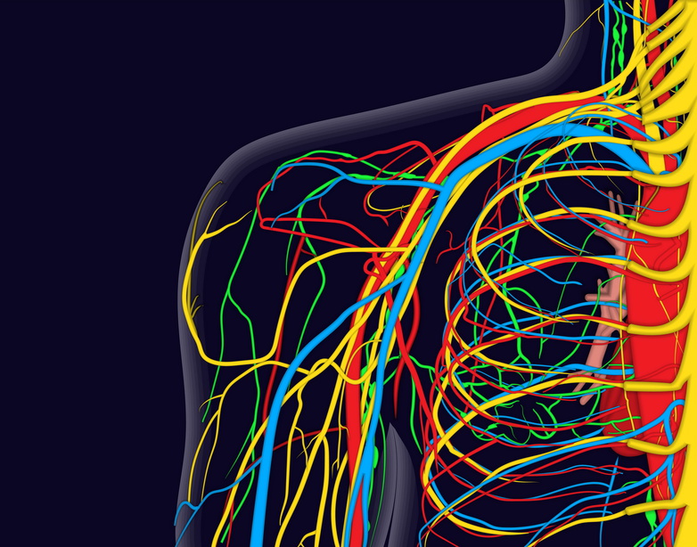
Medical vector illustration of the shoulder anatomy with nerves, veins and arteries, etc.
Cephalic Vein

Image: “Veins of the Arm: Cephalic Vein & Basilic Vein” by Henry Gray. License: Public Domain
Starting around the radial area of what is known as the dorsal venous network, the cephalic vein continues towards the upper part of the body in a circular fashionthroughout the forearm, interacting with tributariesalong the way. Making its way through the basilic vein is the median basilica vein, located in the lower part of the elbow, which works as a communicator in the arm.
Continuing upwards through the forward part of the elbow, the cephalic vein makes its way through the valley created by the Biceps brachii and the Brachioradialis on either side of it. Through this pattern, the cephalic vein comes in contact with the musculocutaneous nerve, continuing up next to the Biceps brachii.
The cephalic vein also travels between the Deltoideuson one side of it, with the Pectoralis major on the other side of it. In this area, joining it is the deltoid branch of the thoracoacromial artery. Next, the cephalic vein travels through the coracoclavicular fascia. It then moves past the axillary artery, completing its journey at the axillary vein, in the area located under the clavicle. The cephalic vein also possesses the capability to interact with the external jugular vein via the clavicle connection.

Image: “Cephalic Vein – Anatomical Dissections” by Anatomist90. License: CC BY-SA 3.0
Accessory Cephalic Vein
Another part of the upper limb that emerges as important is the accessory cephalic vein. It can be found with either the tributary plexus or within the ulnar area making up the dorsal venous part of the upper limb. In some individuals, the cephalic area on the upper side of the wrist is connected to the accessory cephalic. Joining the basilic and cephalic veins through part of the forearm is part of the oblique muscles.
Basilica Vein
Another important upper limb portion to discuss in more detail is the basilica vein. It starts within the ulnar area of the dorsal venous network. It travels a bit in the arm area until it connects with the vena medianacubiti. To meet the vena mediana cubiti, this vein climbs upwards in the back area of the ulnar spot within the forearm, settling in the lower part of the elbow.
Continuing upwards in the valley sides of the Biceps brachii and Pronator teres, the basilica vein passes over the area of the brachial artery. It is kept from connecting with the brachial artery by the lacertusfibrosis. The medial antebrachial cutaneous nerve can also be found on both sides of the basilica vein.
Following this, the basilica vein then continues up, meeting alongside the Biceps brachii’s medial border. Slightly under the middle arm area, it interacts with the deep fascia. The basilica vein then continues down, residing on the bra chart artery’s medial side. It can then be found around the Teres major’s lower area, moving towards the axillary vein area.
Median Antebrachial Vein

Image: “Veins of the Arm – Anatomical dissections” by Anatomist90. License: CC BY-SA 3.0
The next important vein that is part of the upper extremities is the median antebrachial vein. The purpose of this vein is to drain the venous plexus area of the hand as it moves up through the front part of the arm. The median antebrachial vein ends around the basilica vein.
Deep Veins
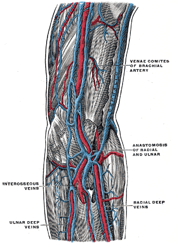
Image: “Deep Veins of the Arm” by Henry Gray. License: Public Domain
There are also deep veins that exist in this area of the body which are important to the arteries and make up the venaecomitantes. Most of the deep veins also come in sets of two, making their homes on either side of the artery. Transverse branches typically join the two veins together.
Within the venaecomitantes are both radian and ulnar veins, with the ulnar veins typically existing as larger in size, while the radial veins interact with the dorsal metacarpal veins. On the other hand, the ulnar veins interact more with tributaries that deal with deep volar venous arches, causing them to have more to do with the wrist area of a human being. This interaction is message sending which follows the profunda vein up into the vena medianacubiti.
Then, on both sides of the brachial artery, one can find the brachial veins. The brachial veins connect with the axillary vein, and the axillary vein then connects with the basilica vein. The deep veins both act with and connect with not only other deep veins, but also with some of the superficial veins as well.
Axillary Vein
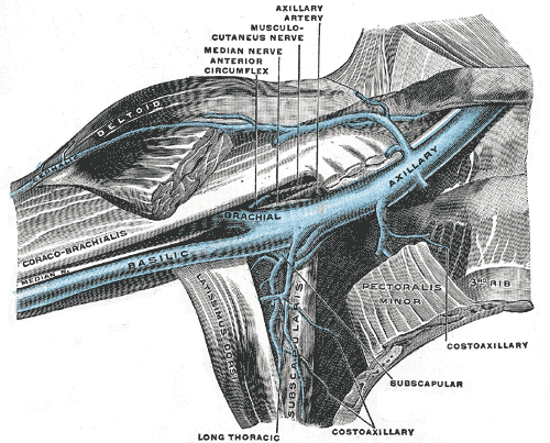
Image: “Anterior view of right upper limb and thorax – axillary vein and the distal part of the basilic vein and cephalic vein” by Henry Gray. License: Public Domain
One of these important veins is the axillary vein; as already discussed, it works with the basilica vein. The axillary vein can be found in the bottom area of the Teres major, getting larger as it progresses upward through the body, completing its journey near the subclavian vein.
The axillary vein has areas that communicate with both the brachial veins and the cephalic vein while at the same time coming near the axillary artery. The axillary vein resides near the axillary artery, following it at times around the brachial plexus near where the medial anterior thoracic nerves are located.
The next important vein is the subclavian vein, which is also known as part of the axillary since it is an extension of it. It can be found around an individual’s first rib, with the other side of it being located near the sternal side of the clavicle. The subclavian vein then meets with the jugular, and this connection makes up what is known as the innominate vein.
To the front of this vein it exists near the clavicle and Subclavius, whereas on the other side of the vein, it sits near the subclavian artery. Two nerves keep this vein apart from the artery: the Scalenus anterior as well as the phrenic nerve. The subclavian vein also is made up of a pair of valves. It can rise upwards through the body as high as the neck.
Cutaneous innervation
Cutaneous nerves that supply the skin of the upper limb are called Dermatomes. Each dermatome is innervated by a spinal cord segment: C3-T3.
Appears to be a confusing pattern. More straight forward if follow the pattern of development.


Comentários
Enviar um comentário