Blood Supply and Venous Drainage of the Brain
Table of Contents
Arterial Blood Supply of the Brain
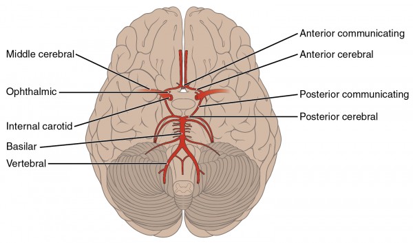
The arterial blood supply of the brain is derived from the vertebral and internal carotid arteries. The vertebral arteries supply blood to the “posterior circulation” and the carotid arteries supply blood to the “anterior circulation”. However, these two important sources of blood are directly connected (think of this as nature’s “back up system”, should one of the systems fail). The connection of the vertebral and carotid circulations is via the Circle of Willis.
Anatomy of the Internal Carotid Artery (Anterior Circulation)
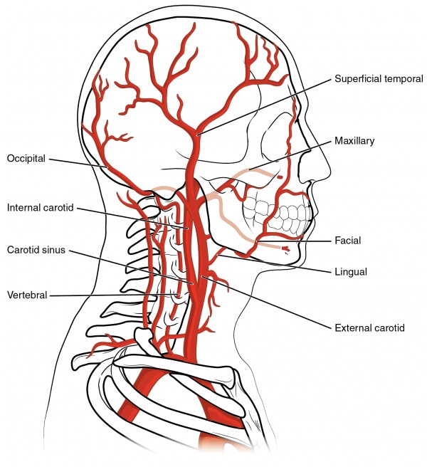
The Common Carotid Artery (CCA) divides at the level of the 4th cervical vertebra (C4) to form the internal and external carotid arteries. This site is referred to as the carotid bifurcation(bifurcation carotidis) and contains the carotid body (glomus caroticum), which contains chemoreceptors that detect changes in the oxygen concentration and the pH-value of blood circulating through the carotid arteries.
The external carotid artery (ECA) divides into eight major branches.
The internal carotid artery (ICA), on the other hand, has no branches in the cervical region. The Internal carotid artery is divided into several segments including: pars cervicalis (cervical segment), pars petrosa (petrous segment), pars cavernous (cavernous segment) and pars cerebral (cerebral segment). Each of these segments, with the exception of the pars cervicalis, has numerous branches. The intracranial portion of the internal carotid artery is further divided into five segments (C1 – C5), terms which are used in clinical practice. The direct continuation of the internal carotid artery is the middle cerebral artery and the anterior cerebral artery, which is the smaller of the two and supplies the medial portion of the hemispheres to the parietal lobe.
The internal carotid artery supplies the frontal, parietal and the temporal lobes, as well as the diencephalon. The ophthalmic artery is a branch of the internal carotid artery, and supplies the eyes and parts of the paranasal sinus.
Vertebrobasilar Circulation (Posterior Circulation)
The vertebral arteries arise from the subclavian arteries and proceed through the transverse foramina (foramina transversaria) of the cervical vertebrae at the levels of C1 through C6. Subsequently, at the level of the foramen magnum, the vertebral arteries pierce the posterior atlantooccipital ligament, the dura mater, and the arachnoid membrane, reaching the subarachnoidal space. The vertebral artery then gives rise to the posterior inferior cerebellar artery, the largest branch of the vertebral artery and one of three arteries that supply the cerebellum.
Another branch of the vertebral artery is the single anterior spinal artery, which is supplied by both vertebral arteries.
In the region of the pons, both vertebral arteries merge to form the single basilar artery. Unlike the vertebral arteries, the basilar artery is an unpaired artery, which means that an occlusion of the basilar artery can lead to devastating clinical consequences. For example, an occlusion of the distal part of the basilar artery can result in an infarction of the pons and the clinical picture of a “locked-in syndrome”.
The basilar artery proceeds along the ventral surface of the pons. Along its course, the basilar artery gives rise to the AICA (anterior inferior cerebellar artery), the pontine perforators, as well as the superior cerebellar artery (SCA). Another possible branch of the basilar artery is the labyrinthine artery, which supplies the inner ear. However, the labyrinthine artery may also arise from the AICA.
Posterior Cerebral Artery
At the transition of pons to mesencephalon, the basilar artery bifurcates into the paired posterior cerebral arteries, one of the three major arteries that supply the brain. There are several branches of the PCA. The posterior communicating artery connects the posterior cerebral artery (and therefore the posterior circulation) to the internal carotid artery (and the anterior circulation). Further important branches of the posterior cerebral artery include the following: anterior and posterior temporal arteries (supply the temporal lobe), medial and lateral occipital arteries (supply the occipital lobe), the calcarine artery (supplies the visual cortex) and the medial and lateral posterior choroidal arteries (which supply the choroid plexus).
The posterior cerebral artery can also be divided into the 4 segments P1 – P4, which is useful to describe the precise localization of a vascular lesion.
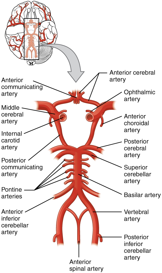
Anatomy of the Circle of Willis
The Circle of Willis represents a circulatory anastomosis between the vascular territory of the carotid arteries, known as the “anterior circulation” and the vascular territory of the vertebrobasilar circulation, also known as the “posterior circulation”. The anterior and posterior circulations are connected at the base of the skull via the paired anterior communicating arteries and the posterior communicating arteries. Therefore, should there be an occlusion in one part of the cerebral circulation, the Circle of Willis allows collateral supply via an alternate vascular route.
The anterior and posterior communicating arteries are also clinically important, because they are a frequent site for intracranial aneurysms (pathologic vascular dilatations), which can result in a subarachnoid hemorrhage should the aneurysm rupture.
It is important to note that there are many normal anatomical variations to the Circle of Willis.
Vascular anatomy of the brain
The middle cerebral artery supplies a large part of the lateral aspect of the hemispheres, including Broca’s area and Wernicke’s areas, the primary motor and sensory language centers respectively. The occipital lobe is supplied by the posterior cerebral artery, and includes the visual cortex. The frontal lobes, medial portion of the hemispheres and the superior portion of the parietal lobes, are supplied primarily by the anterior cerebral artery. For a detailed view of the vascular supply and corresponding anatomy, please review the image.
Note: The arterial vessels run inside the sulci.
Stroke Syndromes
A stroke occurs when there is disruption of cerebral blood flow associated with ischemia in the associated brain tissue. This can occur due to occlusion of a cerebral vessel (ischemic stroke) or due to rupture of a cerebral blood vessel (hemorrhagic stroke). The clinical symptoms and signs of stroke depend on which blood vessel and corresponding brain tissue is affected.
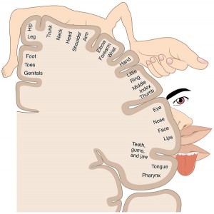
The classical clinical presentation of stroke in the anterior cerebral artery vascular territory includes: hemiparesis (i.e. weakness), involving the legs more so than the arms, always contralateral to the vascular lesion. This is due to the fact that the anterior cerebral artery supplies the frontal, prefrontal and supplementary motor cortex as well as part of the primary motor and sensory cortex. Additionally, occlusion of the anterior cerebral artery can lead to bladder dysfunction. A review of the homunculus may help clarify (please see image).
When the middle cerebral artery territory is affected by as stroke, the classical clinical presentation includes contralateral hemiparesis(weakness), as well as sensory deficits. Aphasia (the inability to speak), may also occur when the dominant hemisphere for language is affected (which is the left hemisphere for most right handed people and many left handed people as well).
Since the posterior cerebral artery supplies the visual cortex, strokes in this vascular territory will typically result in visual disorders, e.g. in the form of hemianopsia. If the vascular supply to the thalamus is also affected, clinical findings can include sensory deficits (numbness) in the contralateral face, arm and leg.
Arterial Supply of the Brain Stem and Cerebellum
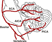
The brain stem and cerebellum are supplied by the basilar artery as well as the cerebellar arteries or branches of these vessels. The branches are differentiated into medial, mediolateral and lateral portions, based on their localization and which anatomical territory they supply. The medulla, which follows the brain stem, is supplied, among others, by the anterior spinal artery, a branch of the vertebral artery.
The cerebellum is supplied by the AICA (anterior inferior cerebellar artery), posterior inferior cerebellar artery (PICA), as well as the superior cerebellar artery (SCA). The AICA is the first branch of the basilar artery. The PICA is a branch of the vertebral artery. The SCA is the final and largest branch of the basilar artery, before it splits into the paired posterior cerebral arteries.
Venous Drainage of the Brain
The venous drainage of the brain occurs via the superficial and deep venous systems, both of which drain via the dural venous sinuses. Both systems are connected via anastomoses. At the transition of both drainage areas, a reversal of the venous flow is possible. Unlike all other veins of the body, cerebral veins do not have valves.
Superficial Venous System
The superficial venous system includes the cortical veins and the sagittal sinus and drains the cerebral cortex. The major cortical veins are named by virtue of their location, and include a superior, middle and inferior group. The superior cerebral veins drain primarily into the superior sagittal sinus and the superficial middle cerebral veins into the cavernous sinus. Based on the direction of the drainage, superficial veins that are located laterally can be further divided into ascending and descending veins.
Ascending veins primarily drain into the superior sagittal sinus, while descending veins prefer to drain into the transverse sinus. Veins that are located between the arachnoid membrane and the dura mater connect the superficial veins with the sinus system, and are also referred to as bridging veins. If these bridging veins rupture, a subdural hemorrhage may occur. The major cause for rupture of bridging veins is trauma.
Deep Venous System
The deep venous system includes the deep cerebral veins, straight sinus, sigmoid sinuses and lateral sinuses. The paired deep cerebral veins and the paired basal veins, are part of the deep venous system.
The basal vein is formed by fusion of anterior cerebral vein and deep middle cerebral vein. Similar to the arterial circulation of the Circle of Willis, there is a venous anastomosis at the cranial base formed by a connection of both basal veins (basal vein of Rosenthal). The anterior communicating vein connects both anterior cerebral veins, which leads to the formation of a closed circulation.
Further venous drainage takes place via the paired deep cerebral veins, as well as the paired basal veins into the Vein of Galen (great cerebral vein). The site where both veins fuse together is also referred to as confluence of sinuses (Torcula) . The great cerebral vein originates at this site.
The great cerebral vein (Vein of Galen) is an unpaired vein and discharges into the straight sinus. The drainage area of the internal cerebral vein includes the thalamus, striatum, choroid plexus, as well as the septum pellucidum. The basal vein drains blood from medial and basal parts of the frontal and temporal lobes, insular cortex and hypothalamus, as well as from the mesencephalon.
Dural Venous Sinuses
The dural venous sinuses are small venous structures that drain blood from cerebral veins, orbits and skull into the internal jugular veins.

Structure of the Dural Venous Sinuses
The dural venous sinuses are located between the periosteal and the meningeal layers of the dura mater, which consists of firm collagenous connective tissue. Besides the connective tissue of the dura mater, the wall of the dural venous sinuses is also made of endothelium. Like all other cerebral veins, the dural venous sinuses do not have valves. The dural venous sinusesshow extensions at some sites, also referred to as lateral venous lacunes. Liquor reabsorption, via the arachnoid villi, takes place in the area of the lateral venous lacunes.
Superficial cerebral veins, the diploic veins of the surrounding periosteum, as well as the emissary veins, discharge into the dural venous sinuses via the bridging veins. The emissary veins connect the dural venous sinuses to extracranial veins, which drain the scalp and onto the diploic veins.
Classification of the Dural Venous Sinuses
Within the system of dural venous sinuses, there is an upper and a lower group. Both groups are connected to the veins of the vertebral canal, via the sinus marginal sinus, as well as the basilar plexus.
The upper group includes superior and inferior sagittal sinuses, occipital occipital sinus, transverse sinus, straight sinus, sigmoid sinus and the confluence of sinuses.
The lower group includes the cavernous sinus, together with anterior and posterior intercavernous sinus, the sphenoparietal sinus, as well as superior and inferior petrosal sinuses.
In – and Outlets of the Dural venous sinuses
Inlets of the dural venous sinuses are superficial, as well as deep cerebral veins. There are numerous anastomoses, which make it possible for larger occlusions in the area of the dural venous sinuses to remain clinically asymptomatic.
The main drainage pathway for the dural venous sinuses is the internal jugular veins. There are also accessory drainage pathways, which include the emissary veins, the superior ophthalmic vein, the marginal sinus and the basilar plexus, as well as the venous plexus of foramen ovale.
Since the cerebral veins and the dural venous sinuses have no valves, the blood flow of the venous system can occur in either direction. This can lead to the spreading of extracranial infections into the sinus system. Infection can further result in an occlusion of a segment of the dural venous sinuses, also referred to as venous sinus thrombosis.
Clinical symptoms of a venous sinus thrombosis include headache, nausea, vomiting and even altered consciousness. These symptoms occur due to an increased intracranial pressure, due to reduced venous drainage.
Additionally, infections can spread via the emissary veins, which form the connection between dural venous sinuses and the extracranial veins, from the scalp to the dura mater, and lead to meningitis.
Venous Drainage of the Brain Stem
The veins in the region of the medulla and brain stem are connected to the basal cerebral veins . The veins of the brain stem are connected to each other via a longitudinal and a transverse network. Furthermore, there is an infratentorial and a supratentorial venous system. Veins arising from the medulla oblongata, pons and cerebellum belong to the infratentorialsystem. The supratentorial system starts at the transition to the mesencephalon. There are numerous anastomoses between the two systems .
Venous Drainage of the Cerebellum
The venous drainage of the cerebellum is differentiated roughly into a medial and a lateral part, with numerous anastomoses between the two parts. The drainage of the cerebellar vermis, the cerebellar hemispheres, as well as the medial part of the superior and inferior cerebellar veins, takes place via the medial system. The remaining parts of the cerebellar hemispheres are drained via the lateral system.
Review Questions
Solutions can be found below the references.
1. Which statement regarding the arterial blood supply of the brain is not true?
- The arterial blood supply of the brain takes place primarily via three large arteries.
- The arterial supply of the cerebellum takes place via three arteries.
- The arterial supply of the brain stem takes place via basilar artery and vertebral arteries.
- Veins and arteries proceed parallel to each other.
- Aphasia can occur if the left middle cerebral artery is occluded.
2. Which statement regarding the Circle of Willis is true?
- The Circle of Willis is a venous anastomosis.
- The Circle of Willis connects the carotid vascular territory with the vertebrobasilar vascular territory.
- The connection at the skull base occurs via middle cerebral artery.
- All aneurysms are located at the posterior communicating artery.
- The structure of the Circle of Willis is the same for all humans.
3. Which statement regarding cerebral veins is not true?
- There are two separate groups of veins, the superficial and the deep veins.
- Cerebral veins do not have valves.
- The dural venous sinuses are venous blood vessels.
- The internal cerebral vein and the basal vein drain into the great cerebral vein.
- Contrary to the arterial system, the venous system has no circulatory bypasses.


Comentários
Enviar um comentário