Hiatal Hernia (Stomach Hernia) — Diagnosis and Treatment
Table of Contents
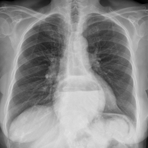
Image: “Large hiatal hernia in the thorax screen” by Hellerhoff. License: CC BY-SA 3.0
Definition of Hiatal Hernia
Hiatal hernia is known as diaphragmatic breakdown
When a hiatal hernia occurs, a portion, or all of the stomach, passes from the abdominal cavity through the hiatus of the diaphragm, intrathoracic.
Classification of various forms of hiatal hernias

Axial hernias (figure C) represent approximately 90 % of all cases and increase significantly in frequency with age: 50 % of all people over 50 years of age have an axial sliding hernia. Here, cardia and gastric fundus relocate into the thoracic cavity. The function of the esophageal sphincter may be limited.
In the so-called paraesophageal hernia (Figure D), part of the stomach (mostly portions of the gastric fundus) with the peritoneal sac pushes through the inguinal ring next to the esophagus (“paraesophageal”). The angle, position of the cardia and function of the esophageal sphincter remain physiological. In severe cases, a complete relocation of the stomach into the thoracic cavity can occur, which is then called an upside-down stomach.
The mixed hernia, a combination of an axial sliding hernia and a paraesophageal hernia, is rare, present in only 5 % of all cases. In up to 40 % of the patients, the hiatal hernia can be accompanied by cholelithiasis. If a sigma diverticulosis is also present, one speaks of the so-called Saint-Trias.
Pathophysiology of Hiatal Hernia
Origin of a hiatal hernia
The esophageal hiatus is an opening located at the level of the 10th to 11th thoracic vertebra. The esophagus, the left and right vagus nerve and portions of the phrenic nerve extend from cranial to caudal into the peritoneal cavity.
The gastroesophageal junction is located within the diaphragm gap (hiatus). Two muscle-faceted muscle strands located centrally on the diaphragm surround the esophagus and form a unit with the esophageal sphincter. In the course of an intra-abdominal pressure increase, a reflux barrier is present physiologically.
The esophageal and gastric fundus should be at an acute angle to each other(about 50° – so-called “His’scher angle“) for optimal barrier function. If there is an expansion of the diaphragm gap, the stomach might shift intrathoracically and the His’sche angle may increase to accommodate the change. Additionally, due to the cranial dislocation of stomach parts, the interaction of the esophageal sphincter and the hiatus is disturbed: Gastroesophageal refluxoccurs.
Symptoms of Hiatal Hernia
Complaints by type of hernia
Sliding hernias are often just an incidental finding and often remain without clinical significance. Due to the limited functionality of the esophageal sphincter, up to 20 % of patients have reflux complaints such as heartburn, bloating or the feeling of pressure and retrosternal pain, especially when lying down. In the course of the reflux, esophagitis can develop, which may be associated with chronic bleeding and thus, an iron deficiency anemia. Rarely, there may be a bolus obstruction when the upper edge of the hernia has narrowed into the so-called schatzki ring (“Steakhouse syndrome”).
Paraesophageal hernias may be symptomatic or show non-specific symptoms such as regurgitation, dysphagia and pressure in the heart area. Reflux symptoms are not typical in these cases, because the esophageal sphincter is indeed intact. However, the paraesophageal hernias involve the risk of incarceration, passage disorders, ulcers and bleeding (iron deficiency anemia!), which is why they are usually operated on while still in the asymptomatic stage.
Diagnostics of Hiatal Hernia
Endoscopy of the hiatal hernia

Image: “Hiatus hernia,” by Adamantios. License: CC BY-SA 3.0
In endoscopy, particularly the so-called “Z-line” is assessed. This line presents itself at the gastroesophageal junction, where the squamous epithelium of the esophagus passes into the columnar epithelium of the stomach. Physiologically, the Z-line should therefore be located in the esophageus hiatus, so exactly where the esophagus passes through the diaphragm.
In a sliding hernia a cranial shift of the Z-line, above the diaphragm occurs as a result of the relocation of the cardia intrathoracically. If the diaphragmatic esophageal passage is less than 3 cm distally to the Z-line, it is known as a small hernia. If there is more than a 3 cm passage from the esophageal hiatus to the Z-line, it is considered a large hernia. Since there will be no reduction of the gastroesophageal junction in the paraesophageal hernia, endoscopically no firm diagnosis on the basis of the Z-line can be made!
Chest X-ray

Image: “Hiatus hernia in lateral radiograph of the thorax. Arrow points at the air-liquid-mirror,” by Hellerhoff. License: CC BY-SA 3.0
In chest X-ray, a possible mirror formation can be detected in the herniated stomach, which is then projected into the thorax and can be substantiated, in accordance with distinct findings.
Barium swallow X-ray
The patient is placed in a head-down position and asked to activate the abdominal prelum. This can provoke a herniation. Constrictions and bottlenecks can be displayed optimally, as well as an intrathoracic shift of the cardia.
Therapy of Hiatal Hernia
Symptomatic or surgical treatment of the hiatal hernia
Axial hernias are treated symptomatically with proton pump inhibitors, if an existing reflux disease is present. Surgical treatment is indicated only in cases of therapy resistance.
Paraesophageal hernias and mixed hernias, however, are always considered a surgical indication, due to the risk of incarceration. Here, the stomach is repositioned and fixed to the anterior abdominal wall, which process is referred to as gastropexy. In most cases, a fundoplication takes place: A fundic wrap, which is sewn around the cardia, serves as a new, mechanical reflux barrier. As part of a hiatoplasty, the diaphragm strands are finally fixed posterior to the esophagus with sutures to narrow the hernia gap.
Review Questions
The correct answers are located below the references.
1. An axial sliding hernia most likely does not present with:
- Pressure or fullness
- Retrosternal pain
- Esophagitis
- Heartburn
- Incarceration
2. Which statement is mostly true for paraesophageal hernias?
- It usually has an asymptomatic course.
- There is a surgical indication.
- Reflux symptoms are in the foreground.
- The cardia of the stomach is usually intrathoracically.
- It is a mixed hernia.
3. Which diagnosis fits the image of a hiatal hernia the least?
- Laboratory: MCV, MCH and ferritin are reduced.
- Endoscopy: the Z-line shifts cranially.
- Chest radiograph: interstitial drawing reproduction.
- Esophagus barium swallow: Imaging of the contrast-filled intrathoracic stomach.
- Increase in reflux symptoms when lying down.


Hernia is commonly occur in stomach. Different types of hernia are also occur in different part of body. This blog also shares valuable information about different types of hernia and diagnose procedure.
ResponderEliminarHernia Specialist Doctor in Delhi Noida | Robotic Metabolic Surgery in Delhi Noida
Thanks for sharing such beautiful information with us. I hope you will share some more information about hiatal hernia. Please keep sharing.
ResponderEliminarHealth Is A Life