Anatomy of the Upper Limb: Muscles of the Hand and Fingers

Hardly any other muscle group has so many small intricate complexes as those of the thumb and fingers and causes such a headache to medical students. And yet, the muscles and their functions can be very easily remembered on the basis of their denominations and the majority of them is innervated by the deep branch. It is, therefore, advisable for aspiring doctors to study with the help of mnemonics in order to be able to go relaxed into their next exam.

Hands and Fingers
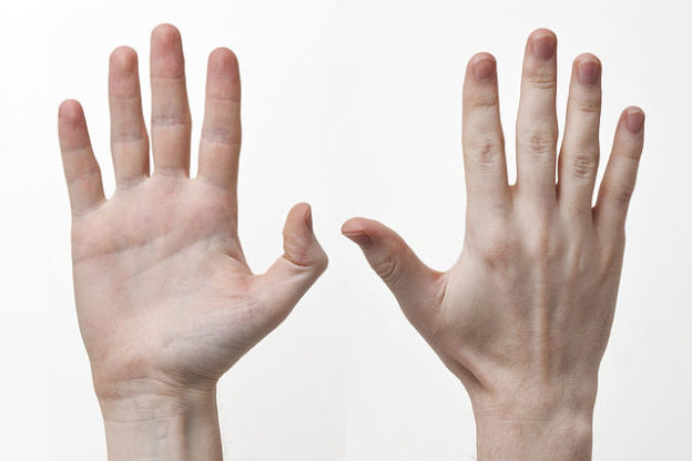
Image : “The front and back of a human right hand” by Evan-Amos. License: CC BY-SA 3.0

diese abbildung zeigt die muskeln die die hand bewegen
Image: “Titel” by philschatz. License: CC BY 4.0
Many of the muscles that move the hand and fingers are reviewed in the Anatomy of the Upper Limb – Muscles of the Arm article.
The other muscles of the hand are divided into 3 groups: the intrinsics, the thenars, and the hypothenars.

Lumbricals

MusclesOriginInsertionNerve supplyFunction
Lumbricals (I-II)Lateral two tendons of FDP (unipennate)Lateral surfaces of extensor expansions of digits  2—5Median nerve (T1)Flex metacarpophalangeal and extend interphalangeal joints of digits 2—5
Lumbricals (III-IV)Medial two tendons of FDP (bipennate)Deep branch of ulnar nerve (T1)
The lumbricals are four narrow muscle bellies which have no direct bony anchoring. They also stabilize the metacarpophalangeal joints and prevent an ulnar deviation.

Interossei Muscles

MuscleOriginInsertionNerve supplyFunction
Dorsal interosseiDorsal sides of all metacarpals (bipennate)Base of proximal phalanges and extensor expansions (digits 2—4, dorsal; digits 2, 4 and 5, palmar)Deep branch of ulnar nerve (T1)Abduct digits 2—4 away from axial line
Palmar interosseiPalmar sides of metacarpals 2, 4 and 5Adduct digits 2, 4 and 5 toward axial line
The radial artery passes through between the two heads of the dorsal interosseous muscle I. There are three palmar interossei muscles and they are one-headed and lie between the metacarpal bones.

Thenar Muscles

MusclesOriginInsertionNerve supplyFunction
Opponens policisFlexor retinaculum and tubercles of scaphoid and trapeziumLateral side of the first metacarpalRecurrent branch of median nerve (C8)Oppose thumb
Abductor pollicis brevisLateral side of proximal phalanx of digit 1Abducts thumb; supports the opposition
Flexor pollicis brevisFlexes thumb
Adductor pollicis Oblique head: Base of second and third metacarpals and capitate
Transverse head: anterior surface of the third matacarpal
Medial side of proximal phalanx of thumbDeep branch of ulnar nerve (C8)Adducts thumb

Hypothenar Muscles

MusclesOriginInsertionNerve supplyFunction
Palmaris brevisTransverse carpal ligament and palmar aponeurosisUlnar palmUlnar nerveWrinkles the skin of the medial palm
Abductor digiti minimiPisiformMedial side of proximal phalanx of the fifth digitDeep branch of ulnar nerve (T1)Abducts the fifth digit
Flexor digiti minimi brevisHook of hamate and flexor retinaculumFlexes proximal phalanx of the fifth digit
Opponens digiti minimiMedial border of the fifth metacarpalRotates and draws anteriorly fifth metacarpal

Review Questions

The solutions can be found below the references.
1. Which of the following structures does not serve as the origin of the extensor digitorum muscle?
  1. Lateral epicondyle humeri
  2. Radial collateral ligament
  3. Os hamatum
  4. Annular ligament of the radius
  5. Antebrachial fascia
2. Which of the following muscles does not belong to the group of thenar muscles?
  1. Abductor pollicis longus muscle
  2. Flexor pollicis brevis muscle
  3. Abductor pollicis brevis muscle
  4. Adductor pollicis muscle
  5. Opponens pollicis muscle
3. Which of the following muscles passes through the 3rd tendon compartment?
  1. Extensor pollicis longus muscle
  2. Extensor pollicis brevis muscle
  3. Abductor pollicis longus muscle
  4. Abductor pollicis brevis muscle
  5. Adductor pollicis muscle

Comentários

Mensagens populares deste blogue

Hemorrhoids (Piles) — Symptoms and Treatment

12 Cranial Nerves — Functions and Mnemonics

Nerve Tissue: Brain