Burns in Children — Classification and Management
Table of Contents
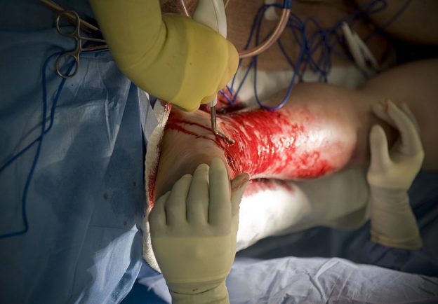
Image: “A burn victim undergoes surgery to remove skin from his legs that will be grafted over his burn.” by U.S. Navy photo – This Image was released by the United States Navy with the ID 090814-N-7090S-040 (next). This tag does not indicate the copyright status of the attached work. A normal copyright tag is still required. See Commons:Licensing for more information. License: Public Domain
Overview
Skin burns in children are common and their management can be challenging due to the difficulty of assessing the depth of the burn in a child.
Classification of Burns in Children
Burns are classified into:
- Superficial burns: a superficial burn is defined as a skin burn that is limited to the epidermis. These burns are usually very painful but can be treated in an outpatient setting.
- Partial thickness burns can be classified into superficial or deep. Superficial partial thickness burns usually involve the epidermis and the dermis and can present with blistering. Deep partial thickness burns usually go deeper than the dermis.
- Full thickness burns are deep burns that are associated with complete destruction of the dermis, hence they are unable to heal on their own. Full thickness burns can also involve deeper structures, such as muscle and bone.
Burns can also be classified by etiology into:
- Thermal burns
- Chemical burns
- Electrical burns
- Frost bits
- Radiation burns
Epidemiology of Burns in Children
Skin burns are common in children and are responsible for approximately 50,000 pediatric emergency visits per year. Approximately, 6,400 children per year are exposed to a serious skin burn that requires admission to a burns unit.
Young children usually sustain skin burns due to scalding, while older children are more likely to be burned by flames or other etiologies.
Pathophysiology of Burns in Children
In contrast to adults, temperatures as low as 40.0°C can cause significant skin damage in young children. The first pathologic change to occur after skin contact with a hot surface is localized capillary stasis and coagulation. This leads to local cellular hypoxia and can cause cellular injury.
Higher temperatures usually cause direct damage to the skin and can destroy the epidermis and/or the dermis. The ability of the burn to heal by re-epithelization is dependent on the presence of a viable dermis. When the dermis is completely or extensively destroyed, the skin loses the ability to heal and skin grafting is usually needed.
Children who acquire a skin burn are at an increased risk of sepsis and extensive fluid loss. The risk of these complications is usually dependent on the depth of the burn in addition to the extent of body surface area involved.
Burns are usually associated with the activation of the stress response which is characterized by the release of pro-inflammatory mediators, hyperglycemia, and a hyperdynamic circulation. Children can develop catabolism and can become feverish. Hyperglycemia can be so severe that insulin therapy might be required even in non-diabetic children.
Burn thickness:
Clinical Presentation of Burns in Children
When a child presents to the emergency department with a burn, the child should be managed as he or she has sustained a trauma. The airway should be assessed to ensure patency, adequate ventilation should be confirmed, and supplemental oxygen administered if patient in distress. The patient’s circulation must be examined, and fluid resuscitation done as per the parklands formula.
Peripheral capillary refill is useful in assessing the peripheral circulation, but the judgment of the extent of fluid loss is better to be based on the extent of the body surface area involved in the burn.
Once the child is deemed to be hemodynamically stable, a detailed history and physical examination should be obtained. The cause of the burn needs to be determined and the possibility of child abuse or neglect needs to be excluded. A local assessment of the burn is useful in determining the depth and extent of the burns.
Taking photographs of the burned areas is recommended for two reasons. The pictures can be shared with other experts and to follow-up the child in the future. Secondly, the pictures can be useful at later stages when abuse or neglect are suspected.
The extent of body surface area (BSA) involved within the burn has to be determined. Subjective assessment is not recommended. The use of charts to assess the body surface area involved is the method of choice in assessing the extent of burns in children. The table below can be used to calculate the percentage of BSA burn in children.
Table 1: Calculation of the percentage of BSA burn in children. Notice that the representing percentage of BSA of the head, thigh and leg differ per the age of the child.
Diagnostic Workup for Burns in Children
The main assessment of a child presenting with a burn should help us answer three main questions. What is the hemodynamic status of the child, what the depth of the burn is, and what is the BSA involved. After adequate fluid resuscitation, the use of strong and adequate analgesia, and securing the airway, breathing and circulation of the child, further diagnostic workup might be carried out.
Certain laboratory investigations might provide some helpful information in the management of the child with burns. A complete blood count can help in the exclusion of anemia, leukocytosis and thrombocytopenia which can complicate a case of severe burn.
Plasma glucose levels should be determined to exclude hyperglycemia. Severe hyperglycemia can be associated with impaired skin healing and should be corrected aggressively.
Urea and electrolytes should be checked at baseline and daily thereafter. After the initiation of fluid replacement therapy. Urine output should be monitored in children who are admitted at a burns unit to assess the adequacy of fluid resuscitation. Urinary catheterization is recommended, and the optimum urinary output should be 1 mL/kg/h.
Treatment of Burns in Children
After assessing the depth and extent of burns in a child, the main next step in the management should be adequate fluid resuscitation. Immediate cooling is recommended in children with burns, but should not be prolonged if the BSA burn is more than 15 % to avoid hypothermia.
Fluid replacement therapy should be initiated as early as possible. For the first 24 hours after the burn, the Parkland formula should be used. The Hartmann’s solution should be used whenever available.
Parkland formula:
V = 4 x m x (A x 100)
The total resuscitation amount of fluid needed per Parkland formula can be calculated by multiplying 4 mL of Hartmann’s solution by the child’s weight by the calculated percentage of BSA burn. The first half of the calculated amount should be given within the first 8 hours after the burn, while the remainder half should be given in the subsequent 16 hours.
After the initial resuscitation, maintenance fluid therapy should be given during the admission and oral intake should be encouraged to be established as soon as possible. The following formula can be used to calculate the amount of maintenance fluid needed for the child: 4 mL per kg per h for the first 10 kg of body weight + 2 mL per kg per h for the next 10 kg of body weight plus 1 mL per kg per h for the remainder of the weight of the child. Hartmann’s solution should be used whenever available.
Children who will undergo surgical skin grafting should undergo general anesthesia by intravenous induction whenever possible. If not possible, then inhalation induction can be used. Succinylcholine should be avoided in children with significant skin burns because of the increased risk of fatal hyperkalemia.
Adequate analgesia should be used in any child with skin burns. Pain can be intense and is better to be based on self-measurement of pain in a cooperative child. Paracetamol and non-steroidal anti-inflammatory drugs can be used to manage pain, but they are usually not enough. Opioids are usually needed in the management of pain in children with significant skin burns but care must be taken to avoid unwanted side-effects, such as respiratory depression, tolerance or dependence.
Non-steroidal anti-inflammatory drugs are usually used in small doses because of the risk of participating or exacerbating kidney injury in a child with burns. COX-2 antagonists are not recommended in the management of pain in children with burns.
Ketamine is better used as an analgesia during dressing changing and bathing and not as a regular pain killer due to its association with delirium. Chronic pain after healing can be treated with gabapentin or pregabalin.
Children with significant pain can become depressed. Play therapy and distraction techniques should be used to avoid psychological trauma to the child.
Children who have a rebound fever, a skin rash, or who become leukopenic should be evaluated for the possibility of toxic shock syndrome. Children can also present with shock, vomiting or diarrhea if they develop toxic shock syndrome. Toxic shock syndrome requires immediate hospitalization if the child was already discharged, adequate fluid resuscitation and the administration of intravenous antibiotics.


Comentários
Enviar um comentário