Hereditariedade e Aconselhamento Genético
Table of Contents
What is Genetic Counseling?
Genetic counseling is the process through which knowledge on the genetic aspects of illnesses is shared by trained professionals with those at an increased risk of having an inheritable condition or passing it to their offspring. It involves a genetic counsellor who talks to the patient about inheritance of an illness and their recurrence risks.
Counseling is aimed at diagnostic purposes, at detecting causes, or at making prognoses about genetic diseases, e.g., through pedigree creation or molecular genetic testing. It is of the utmost importance that the physician provides information but does not influence the patient’s opinion, nor steer his or her decisions in a certain direction. This non-directivity preserves the patient’s autonomy.
Creation and Analysis of Family Pedigrees
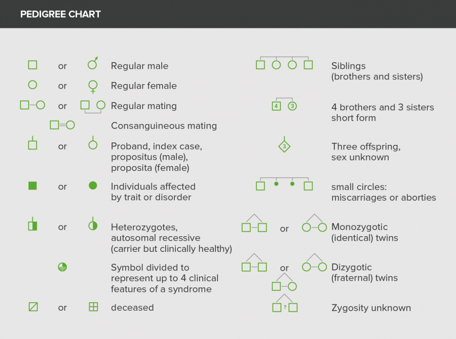
A pedigree (or family tree) is used for a clear presentation of family history, and thus helps to identify abnormalities and inheritance patterns. A pedigree chart shows the occurrence and appearance of phenotypes of a particular gene among family members and their ancestors. First step in creating a pedigree is consulting with the patient and systematically questioning him/her about parents, siblings, children, etc.
Mostly, 3 generations are sufficient to assure reliable results. Inquiries relate to names, birthday and clinically relevant information. Provenance, miscarriages, or unwanted childlessness can also be of interest. The symbols are the same in almost all pedigrees.
Preliminaries: Mendelian Genetics
For a meaningful interpretation, one has to be familiar with the basic rules of Mendelian genetics. The rules are based on the premise that genetic properties are passed on from one generation to the next following a distinct system.
The rules are as follows:
Law of Dominance: Descendants of opposite homozygous parents are all uniformly heterozygous. Not all alleles are phenotypically expressed and when two different alleles are inherited then, one determines the phenotypic characteristics of the organism. This is known as the dominant allele whereas the other is silent and thus it is known as the recessive allele.
Law of Segregation: Descendants of heterozygotes are not uniform, the phenotypes, however, split in a certain ratio during the production of gametes.
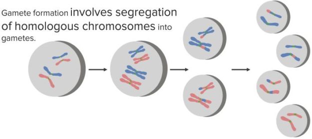
“Homologous chromosomes (alleles) separate during Meiosis” Image created by Lecturio
Law of Independent Assortment: Different characteristics are distributed independently in the next generation.
For each characteristic, we have two alleles, i.e. two responsible genes. One is inherited from the mother, one from the father. If the alleles are on an autosome, it is called an autosomal inheritance. If they are on a sex chromosome, it is a gonosomal inheritance and a number of particularities must be taken into account.
If both alleles have the same effect, the genotype (i.e. the combination of genes) is homozygous, if they differ, then it is heterozygous. The phenotype (the physical appearance of the genetic trait) depends on the type of inheritance.
Different alleles are often represented with upper and lower case letters. A trait can have the gene variation “A” and the gene variation “a”. A homozygous genotype is then represented by “AA” or “aa”, a heterozygous genotype by “Aa”. The capital letter stands in most cases for the dominant allele, the lower case letter for the recessive allele.
Modes of Inheritance
There are 8 classifications of modes of inheritance.
1. Co-dominant inheritance
In case of heterozygous genotypes, both characteristics are physically manifested in parallel. An example of that is the AB0 system of blood types. In a genetic heterozygote AB, both A and B characteristics are phenotypically expressed on the erythrocytes’ surface.
2. Intermediate inheritance
In case of a heterozygous genotype, both characteristics are manifested but not in parallel. Instead, there is an intermediate phenotype, i.e. a blending of both characteristics. The simplest example is the color of a flower: a red allele and a white allele result in a pink colored plant.
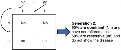
3. Autosomal dominant inheritance
One allele is dominant to another allele. If there is a dominant and a non-dominant allele in a heterozygous genotype, only the dominant allele is manifested. In genetic counselling this represents the fact that in pregnancy there is a 50% chance that the fetus will acquire the disease. It is often referred to as vertical inheritance as the transmission is from the parent to the offspring. Taking the AB0 system as an example again: A and B are each dominant to 0. Therefore, the genotype A0 has the phenotype A.
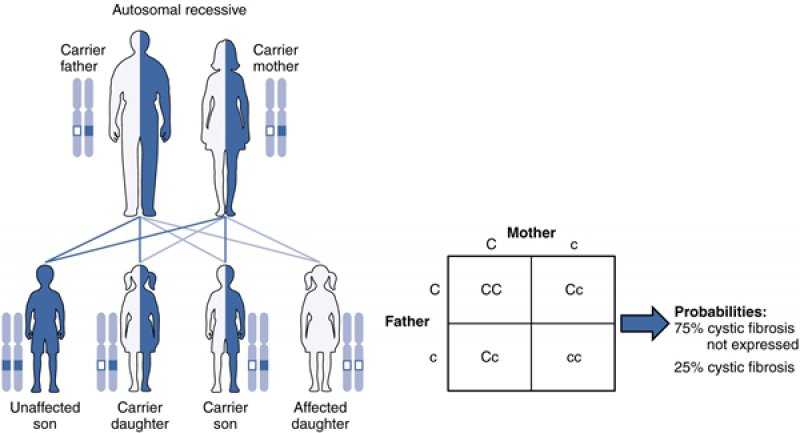
4. Autosomal recessive inheritance
Recessive alleles are only manifested when they are homozygous. The blood type 0 is therefore only possible if the genotype is 00. There are many people with heterozygous genotypes that are not physically manifested.Two copies of an allele are required to phenotypically express the disease.Diseases with this kind of inheritance include sickle cell disease and cystic fibrosis. They are observed in consanguineous relationships as these people have an increased risk of having identical genetic mutations.
5. X-linked recessive inheritance
The allele in question, or the disease-causing mutation, is on the X chromosome and isrecessive. In a male, the allele will always be manifested, because he possesses only one X chromosome. In a woman, the likelihood of manifestation is very low, since she possesses a second X chromosome. Only if there is, for various possible reasons, a non-randominactivation of the healthy X chromosome, the trait will be manifested. For the children of a hemizygous healthy man and a woman, heterozygous for a X-linked recessive genetic disease, the following will apply:
- The male offspring have a risk of developing the disease of 50 %.
- The female offspring have a risk of disease of nearly 0 %; however, there is a risk of 50 %that they are carriers.
If the man is hemizygous for the mutated allele, all male offspring will be healthy (the X chromosome comes from the mother!) and all daughters are healthy female carriers (their second X chromosome is from the father, which is present only in its mutated form!).
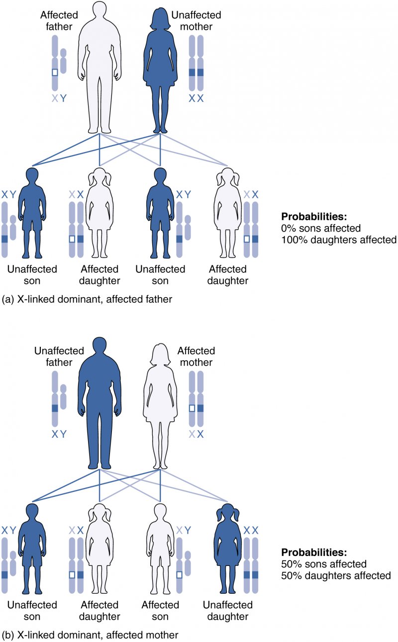
6. X-linked dominant inheritance
Only one copy of the disease allele is required for phenotypic expression as in the autosomal dominant inheritance. However, for the x-linked dominant inheritance the disease allele is located on the X chromosome.This type of mutation is often fatal in male offspring, since they are hemizygous, and the changes are severe. In heterozygous women, the disease also manifests but is attenuated by the second X chromosome. According to the Lyon hypothesis, the female body consists of a mosaic of cells where either one or the other X chromosome is activated.
7. Y-linked inheritance
In case of a holandric inheritance, a diseased father passes on the mutation to all his sons, who will also be ill. However, there are hardly any known diseases with this pattern of inheritance.
8. Mitochondrial inheritance
Also genetic defects of the mitochondrion can cause diseases. Mitochondria are always inherited from the mother. All of the mother’s children can be affected, whereas a father with this type of genetic mutation cannot transmit it.
Definition: Carrier Status
If a person possesses a heterozygous genotype for a recessive hereditary disease, he or she is a carrier. This means, the disease does not manifest, since he or she also has a non-mutated gene. However, he/she can pass it on to offspring, so that the disease occurs in the next or next-but-one generation. Thus, he/she is the carrier of the hereditary disease. For some diseases, a slight pathology can be observed in carriers; however, this varies among individuals.
Case Study: Cystic Fibrosis
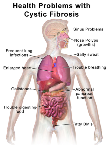
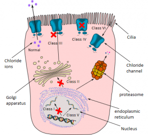
Cystic fibrosis, also called mucoviscidosis, is an autosomal recessive hereditary disease. Cause is a chloride channel that is not functioning properly due to the mutation or is missing completely. Therefore, the composition of different secretions in the body is significantly changed.
Distribution of the Gene Defect
Cystic fibrosis is one of the most common metabolic diseases in our population. It has an incidence of 1:2500, one in twenty Northern Europeans is a carrier. This means, the probability of two heterozygous parents is 1:400, the probability for them to have a diseased child is 1:1600.
Pathogenesis: Symptoms of Cystic Fibrosis
- Viscous mucus in the lungs that causes lung damages and increased risk of infections
- Meconium ileus
- Pancreatic insufficiency of the exocrine pancreas
- Increased chloride and sodium concentrations in sweat
Genetic counseling
The following example is taken from the textbook “New Clinical Genetics” in a shortened form.
David and Pauline already have a 4-year-old healthy boy, when they have another child, a girl named Joanne. Since her birth, Joanne has hardly gained weight. She was often suffering from colds and coughs. At 5 months, she develops a severe pneumonia and is hospitalized. The staff notes the voluminous, foul-smelling bowel movement and the lack of development, since she is now on lower percentiles than at her birth.
Suspected cystic fibrosis is confirmed by a sweat test. With the help of a geneticist, a pedigree is created which, however, identifies Joanne as the only diseased family member. This is a typical presentation for autosomal recessive diseases, since a single mutation on one allele only results in inconspicuous carriers. In order to clarify the exact type of mutation Joanne has, a molecular genetic test is conducted. This is especially important for relatives who might also be carriers. They can get tested for the corresponding mutation.
Molecular Genetics of Cystic Fibrosis
The autosomal recessive disease is triggered through a mutation of the CFTR gene on chromosome 7 (exact location: 7q31.2). With regard to the defective protein of the patient, it is a homozygous gene defect, since both genes are mutated and, thus, the protein is inactive.
However, this does not mean that the patient has the same mutations on both alleles. There are approximately 1000 identified mutations; determining the disease’s progression is the weaker mutation. This state is called compound heterozygosity. Among these 1000 mutations, there are only a few that occur frequently. The most important mutation is the p.Phe508del, which is responsible for 70-80 % of all cases in Northern Europe.
Joanne has been tested for the most common mutations by PCR. She is heterozygous regarding the mutation p.Phe508del. This means she has a second, yet unknown mutation. Through further testing, a mutation is identified that her mother has as well and which has been found before in other cystic fibrosis patients.
Joanne’s relatives can now get tested for these two mutations.
Review Questions
Solutions can be found below the reference.
1. Which mode of inheritance shows no vertical inheritance pattern?
- Autosomal recessive
- X-linked dominant
- Autosomal dominant
- Mitochondrial
- Y-linked
2. Which statement about cystic fibrosis is true?
- Cystic fibrosis is always caused by the mutation p.Phe508del.
- One in four Europeans is carrier of this disease.
- Symptoms are caused by the lack of a chloride channel.
- The disease is triggered only a few months after birth.
- The children of two carriers are always affected.
3. What is true for carriers?
- They are often found in autosomal dominant diseases.
- The risk for girls to be the carrier of a X-linked recessive disease is 50 %.
- The risk for girls to be the carrier of a X-linked recessive disease is 100 %.
- The risk for girls to be the carrier of a X-linked recessive diseases is 25 %.
- The risk of disease in case of one parent with autosomal dominant disease is 100 %.

Comentários
Enviar um comentário