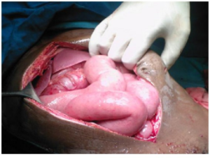
Abdominal and Pelvic Injuries: Blunt and Penetrating Abdominal Traumas (Gunshot Wounds and more)
Table of Contents
Image: “Various cooking knives.” by WLU – Own work. License: Public Domain
Epidemiology of Abdominal and Pelvic Injuries
World over, firearm mortality rates have varied across different regions owing to laws governing the use of firearms, from 0.05 in Japan to 14.24 in the USA.
- In blunt injuries, the solid organs liver and spleen are the most commonly injured organs.
- In penetrating injuries, however, one-half of the patients have their small bowels injured followed by colon, liver and vascular structures.
Mechanism of Injury
It is helpful to know the anatomical regions of the abdomen and its contents for a better understanding of the mechanisms underlying the abdominal and pelvic injuries.
There are three distinct compartments namely:
- Peritoneal cavity: Subdivided into the intrathoracic and abdominal segment. The intrathoracic segment is covered by bony thorax, which includes diaphragm, liver, spleen, stomach, and transverse colon.
- Retroperitoneum: This is a particularly difficult compartment because of its remote location rendering it less yielding towards a physical examination and peritoneal lavage. This region houses the aorta, vena cava, pancreas, kidney, ureters, and portions of duodenum and colon.
- Pelvic compartment: because of its anatomical location, injuries to the rectum, bladder, iliac vessels, and internal genitalia of women are difficult to diagnose.
Abdomen and pelvic trauma can occur due to blunt, penetrating, and explosion forces. In polytraumatic cases, blunt trauma forms the major bulk while penetrating injuries are less common.
Blunt injuries
Three mechanisms explain the occurrence of blunt injuries:
- Deceleration: A differential movement between adjacent structures owing to a rapid deceleration lies at the core of this mechanism. Shear forces cause hollow, solid organs and vascular pedicles to tear along their lines and points of attachment. So in classic injuries of the liver, tears along ligamentum teres are often encountered, as are mesenteric tears along bowel loops injuring the splanchnic vessels as well.
- Crushing: the solid organs trapped between the anterior abdominal wall and posterior thoracic cage are susceptible to crushing injuries.
- External compression: The compression could be a result of a direct blow or from external compression against a rigid fixed structure. In accordance with Boyle’s law, hollow organs are especially vulnerable as the compressive forces result in a sudden rise in intra-abdominal pressure.
Penetrating injuries
- Gunshot wounds: While a gunshot wound involves a high energy transfer giving rise to an unpredictable pattern of injuries, additional damage is done by bullet and bone fragments. The severity of a shotgun varies according to the distance of the victim from the weapon.
- Stab wounds: stab wounds have a more predictable pattern of injury wherein penetration of the abdominal wall is caused by a sharp object.
In both types of penetrating injuries, the mode of injury determines the underlying mechanism. While homicide is the predominant mode of injury in the adult population, children are more susceptible to accidental penetrating injuries at home.
Clinical History and Examination
A detailed history and careful examination remain the cornerstone of management of patients with abdominal and pelvic trauma.
- Primary survey includes assessment and concurrent resuscitation by following the ABCDE protocol. After initial resuscitation, a level of consciousness / Glasgow Coma Score (GCS) is assessed.
- The patient should be completely undressed for a complete head to toe examinationincluding the back and perineum areas that are often missed out but are sources of significant bleeding.
- Abdominal distension, tenderness, obliterated liver dullness, deformed pelvis with tenderness are all suggestive of an intra-abdominal and/or pelvic injury.
- Relevant investigations should be performed to confirm clinical suspicion.
- Patients with altered mental status or pelvic and retroperitoneal organ injury make examination difficult and are potential candidates for investigations.
Investigations
Diagnostic tools in care of trauma patients have a specific role in:
- Confirmation of clinical suspicion
- A decision regarding nature of procedure to be performed
- Evaluating and monitoring patient receiving nonoperative treatment
Chest X-ray (Antero-Posterior and Lateral views)
- A chest x-ray is most often used to rule out chest involvement. It also provides information regarding free intraperitoneal gas, herniation of abdominal contents etc.
X-ray Pelvis
- To rule out pelvic bone fracture as a source of bleeding.
FAST (Focused Assessment with Sonography for Trauma)
In patients with polytrauma who are hemodynamically unstable and refractory to fluid administration and blood transfusion, bedside FAST is the only imaging method beneficial to them. Free fluid in the abdomen and pelvis suggest intraabdominal hemorrhage.
- Sensitivity: 73-88 %
- Specificity: 98-100 %
Drawbacks:
- Grading of solid organ injury not possible
- Mild hemorrhage may be missed
- May cause injury to retroperitoneum
- Operator dependent
CT Scan Abdomen with Contrast
It is the investigation of choice for hemodynamically stable patients.
- Provides information regarding injury to retroperitoneal structures, diaphragm, and solid abdominal organs.
- Grading of injuries can be obtained.
- Presence of free fluid in the abdomen in the absence of solid organ injury is suggestive of bowel, mesenteric or urinary tract injuries.
- Accuracy: 92-95 %
- Highly sensitive and specific for hepatic and splenic injuries
Diagnostic peritoneal lavage (DPL)
- When encountered with blunt trauma, DPL is reserved for patients with spinal cord injury or multiple injuries with unexplained shock, especially if the mental status of the patient is altered. Intoxicated patients with suspicion of abdominal injuries or those who will undergo prolonged anesthesia are other candidates.
- A viable alternative in institutes where CT scan and FAST facilities are not available.
- It is an invasive procedure wherein a soft catheter is introduced into the peritoneal cavity for aspiration of lavage fluid or content that is then evaluated.
- Accuracy: 92-98 %; considered to be more accurate than CECT in early diagnosis of bowel and mesenteric injuries.
- Operative intervention indicated when:
- At least 10 ml of free-flowing blood is aspirated
- Presence of:
- RBC > 100,000/mm3
- Leukocyte count > 500/mm3
- Amylase level >175 U/dL
- Bile, bacteria or food particle
Drawbacks:
- Procedural difficulty in performing in patients with prior surgeries
- Pregnant women
- Obesity
Specific Types of Injuries
Splenic injury
Clinical presentation
- A detailed history regarding the anatomical location of the injury gives a clue to splenic damage.
- Lower left rib fractures must not be ignored as they are often associated with splenic rupture.
- Severe chest or neurological damage make an assessment of minor splenic trauma difficult.
- History of malaria, lymphoma, hemolytic anemia is important as even minor trauma can cause disproportionate damage to an enlarged spleen.
Investigations
- While all basic laboratory tests may be performed, a complete blood cell count is the most useful investigation in pointing towards deteriorating hemodynamic stability.
- The most specific and sensitive study for splenic injury is CT scan abdomen (triple helical scan). It is up to 98 % sensitive for splenic injuries when IV contrast is given. While it can detect small quantities of blood in the abdominal cavity, it is contraindicated in hemodynamically unstable patients.
- Other investigations include FAST, diagnostic peritoneal lavage, and angiography but these have a limited role to play.
Management
- Immediate splenectomy indicated in:
- Patients with severe multiple injuries
- Splenic avulsion
- Fragmentation or rupture
- Extensive hilar injuries
- Failure of hemostasis
- Peritoneal contamination from gastrointestinal tract
All patients are to be administered polyvalent pneumococcal vaccine post-splenectomy.
- Conservative approach
- In patients younger than 55 years with no other associated abdominal injuries, a conservative approach may be used given their hemodynamic stability.
- Patients are kept under an observation period for 10-14 days; delayed rupture and hemorrhage may occur, usually in first 48 hours. This is followed by bed rest for a week.
- They are advised to not indulge in any strenuous activity for a period of 6-8 weeks and engage in sports activities for a period of 6 months at least.
Hepatic injury
It is quite a common injury that is encountered in the emergency department, yet also the most frequently missed injuries in trauma deaths. An abdominal examination may provide vague clues to an injury but these are often missed. For this reason, diagnosis is usually made at laparotomy or CT scan.
Investigations
Basic laboratory tests:
- Complete blood cell count (CBC)
- Coagulation profile: Along with CBC, these two basic lab tests are done to obtain baseline levels of PT, APTT and platelet count as dilutional coagulopathy and thrombocytopenia are common after hepatic repair.
- Clotting factors
- Liver and kidney function tests
- Electrolytes
Imaging studies
- Chest X-ray to rule out chest involvement
- CT scan abdomen is considered to be the most specific and sensitive test for liver injury.
- Diagnostic laparoscopy: since an associated injury to the diaphragm is common with hepatic injury, a diagnostic laparoscopy makes for a good test for ruling out such associated injuries.
- Angiography is less valuable as a diagnostic tool but transcatheter embolization has found use in the management of persistent hepatic bleeding that is not stopped by surgery.
Management
- Packing and limited surgery is a viable option when coagulopathy and hypothermiadevelop.
- Conservative management is more often used for hemodynamically stable patients with no other associated abdominal injuries and peritoneal signs.
Pelvic injury
It is caused by high energy blunt trauma following a fall, road traffic accident or crush injury. Pelvic injuries account for 13-23% mortality.
Clinical features
- The patient presents with a typical history of pain on movement and gross hematuria.
- A quick observation reveals structural instability with peri-pelvic ecchymoses.
- A digital rectal examination is important to identify injury to the rectum and locate the prostate.
- Hemoperitoneum may lead to hypotension.
Investigations
Basic laboratory tests:
- Complete blood cell count has an important role in the management of pelvic bleeding.
- Urinalysis
- Electrolytes
- Liver and kidney function tests
- Coagulation profile
Imaging studies
- X-ray to rule out pelvic bony fractures and associated abdominal injuries
- Diagnostic peritoneal lavage performed at the earliest in hemodynamically unstable patients as it is a good modality to identify hemoperitoneum.
- Laparotomy: if gross bleeding is seen, this should be followed by external fixation and angiography.
Management
The main aim of management of pelvic injury is assessment and control of bleeding.
- Hemodynamically unstable patient: Early open DPL should be performed
- Gross bleeding on laparotomy: External fixation helps to minimize bleeding from veins and small arterioles near fracture sites. It also adds to the tamponade effect by shrinking the volume of an open pelvic cavity. This is followed by angiography with embolization, which is often effective in controlling arterial bleeding but is difficult to perform.
- Large vessel bleeding: Surgical control
- If bleeding is hinted only by low blood cell counts, a risk of major intraabdominal hemorrhage is low.
Penetrating injuries
Clinical presentation
- A detailed history regarding the anatomical location of the wound and the type of weapon used is important.
- A number of gunshots heard or a number of times the patient was stabbed, the position of the victim and the environment under which the incident took place help assess the severity and extent of the damage.
- Additional information regarding allergies, current medications, history of any prior illness or surgery, and last meal had by the victim help in providing the best management possible.
Signs and symptoms
- Primary survey: Initial examination for assessment of ABCDE should be done in the emergency department:
- ABC – Airway, Breathing, and Circulation
- D – Disability: Level of consciousness and neurological deficits should be assessed
- E – Exposure: Inspect all body surfaces for wounds and injuries
- Amount of blood loss
- Type of weapon or object used
- Secondary survey includes complete head to toe examination (in hemodynamically stable patients).
- A rapid abdomen examination for distension, dullness to percussion, and bowel sounds coupled with a digital rectal and genitourinary examination should be done.
- In life-threatening cases, a secondary survey may be reserved for after the operative therapy.
- Indications for immediate surgical exploration:
- Hypotension
- Narrow pulse pressure
- Tachycardia
- High or low respiratory rate
- Peritoneal signs (pain, guarding, rebound tenderness)
- Diffuse or poorly localized pain that fails to resolve
Investigations
- Basic laboratory tests for patients undergoing immediate surgery:
- Blood type and crossmatch
- Complete blood cell count
- Liver and kidney function tests
- Blood glucose
- Coagulation profile
- Arterial blood gas
- Urinalysis
- Electrolyte levels along with calcium, phosphate and magnesium levels
- Toxicology screen
- Imaging studies:
- Chest X-ray: Initial investigation to rule out involvement of chest cavity
- Abdominal radiography: Both anteroposterior and lateral views taken
- Chest and abdominal ultrasonography: Focused assessment with sonography for trauma (FAST)
- CT scan abdomen: Most sensitive and specific study in cases of liver or spleen injury
Management of Abdominal and Pelvic Injuries
A multi-faceted approach is used to manage the patient, which can be described under the following headings:
Diagnostic/therapeutic procedures
These include procedures that are used for monitoring the status of the patient and also help in the management of the patient.
- Gastric decompression
- Foley catheterization
- Peritoneal Lavage
- Tube thoracostomy
- Local wound exploration
Surgical management
- Gunshot wounds are almost always associated with intraabdominal injuries that mandate laparotomy.
- Stab wounds, in contrast, have a lower incidence of intraabdominal injuries and hemodynamically stable patients may be provided expectant treatment.
Medical management
It is very much palliative in nature with obvious choices:
- Analgesics: morphine, fentanyl
- Anxiolytics: lorazepam, midazolam
- Antibiotics: metronidazole, gentamicin, vancomycin
- Neuromuscular blocking agents: succinylcholine
- Immune enhancement: tetanus toxoid
- IV fluids


Comentários
Enviar um comentário