Cytology: Structure and Functions of the Cell
Table of Contents
- Definition of a Cell
- Eukaryotic and Prokaryotic Cells in Comparison
- Structure and Function of the Cell Membrane
- Structure and Function of the Nucleus
- Functions of the Cytoplasm
- Classification of Cell Organelles
- Structure and Function of Individual Cell Organelles
- Components and Functions of the Cytoskeleton
- Structure and Function of Cell Contacts
- Cell Communication
- Review Questions
- References
Image : “Stem Cell” by
PublicDomainPictures. License: Public Domain
Definition of a Cell
The cell as a biological organizational unit, is the smallest basic element of all organisms. It is autonomous and fulfils basic essential functions in metabolism, growth, movement, reproduction and heredity.
Eukaryotic and Prokaryotic Cells in Comparison
Eukaryotic cells, also called eucytes, are 10-100 µm in size and possess a nucleus that contains the DNA of several chromosomes. In addition to exons (coding DNA) the DNA comprises of many introns (non-coding genes) that are removed by processes such as splicing, through protein biosynthesis.
The cytoplasm is heavily compartmentalized and is rich in cell organelles. Ribosomes have a molecular mass of 80S for 60S and 40S subunits (amount of mass as Svedberg centrifugation constant). The respiratory chain takes place in the mitochondria. Examples of Eukaryotes are fungi and animals (from worms to humans).
A prokaryotic cell, also called protocyte or procyte, however, has a size of only 1-10 µm and contains a nucleus equivalent (nucleoid) instead of a nucleus. This “nucleus-like” densely packed molecule is located in the cytoplasm and comprises the DNA, which encompasses of only one chromosome and exhibits no introns.
In addition, a plasmid may be present (circular, extrachromosomal DNA), which plays a special role in the development of antibiotic resistant bacteria. The cytoplasm is less compartmentalized and the respiratory chain is specifically located in the cytoplasm membrane.
While mitochondria, Golgi apparatus and the endoplasmic reticulum are lacking, the ribosomes have a molecular mass of 70S for 50S and 30S subunits. Bacteria such as Escherichia coli belong to the prokaryotes group.
These differences are common exam topics in biology as well as in biochemistry.
Structure and Function of the Cell Membrane
The cell membrane, also called plasmalemma, confines in the cytoplasm and serves as the delimitation between intra- and extracellular space. It is composed of a phospholipid bilayer, whereby the hydrophilic parts of the phospholipids are directed to the intra- and extra-cellular space. The hydrophobic parts are centrally located within the membrane.

The peripheral glycocalyx consists of sugar chains (polysaccharides) that are covalently bounded to membrane proteins (glycoproteins) and membrane lipids (glycolipids). It is individual and cell type-specific, this means for example, that it determines the blood group characteristics of the erythrocytes.
Facilitated by its fluidity, the cell membrane is stable and flexible at the same time. Its fluidity can change depending on temperature and lipid composition. The membrane is semi-permeable (also referred to as selective permeability), which means it is well permeable for small-molecular substances like water, which are able to diffuse osmotically. Higher molecular substances such as proteins require specific transport systems in order to by-pass the cell membrane.
The cell membrane’s functionality is decisively determined by its membrane proteins, which include: ion channels, cell adhesion molecules, aquaporins, membrane pumps, carrier proteins and receptor proteins.

Structure and Function of the Nucleus
The cell core (nucleus) contains DNA, packed in chromosomes, and can vary in both size and structure depending on its activity state. The karyoplasm is separated from the cytoplasm by the porous nuclear membrane, karyolemma.
The nuclear membrane consists of an outer and inner nuclear membrane and the space in between, the perinuclear cistern. The outer nuclear membrane merges into the endoplasmic reticulum and is occupied by ribosomes. The inner nuclear membrane is inside the felt-like nuclear lamina (lamina nuclearis) that is formed by a 30-100 nm sized layer of intermediate filaments.
About 1000-4000 nuclear pores ensure the exchange of substances between the cytoplasm and the karyoplasm, whereby molecules < 5 kDa diffuse freely and larger molecules such as those of proteins pass through by means of receptor binding.
The nucleus contains a small spherical nucleole (nucleolus), from which the ribosomal RNA originates. The transcription as a prerequisite for translation as well as the replication as a prerequisite for mitosis is also controlled by the nucleus.

Functions of the Cytoplasm
The cytoplasm, also called cytosol, is restricted by the cell membrane and represents the fluid matrix of each cell. The cytoskeleton, cell organelles as well as cell inclusions are embedded in the cytoplasm.
Within the cytoplasm protein biosynthesis, ion currents as well as vesicle transport takes place around the Golgi apparatus, the endoplasmic reticulum and the cell membrane. It constitutes about 50 % of the cell’s volume and has a pH of 7.2.
Classification of Cell Organelles
Cell organelles are embedded in the cytoplasm and are divided into:
- Membrane-restricted organelles (rough and smooth endoplasmic reticulum, Golgi apparatus, mitochondria, lysosomes, peroxisomes)
- Non-membrane-restricted organelles such as ribosomes or centrioles
Structure and Function of Individual Cell Organelles
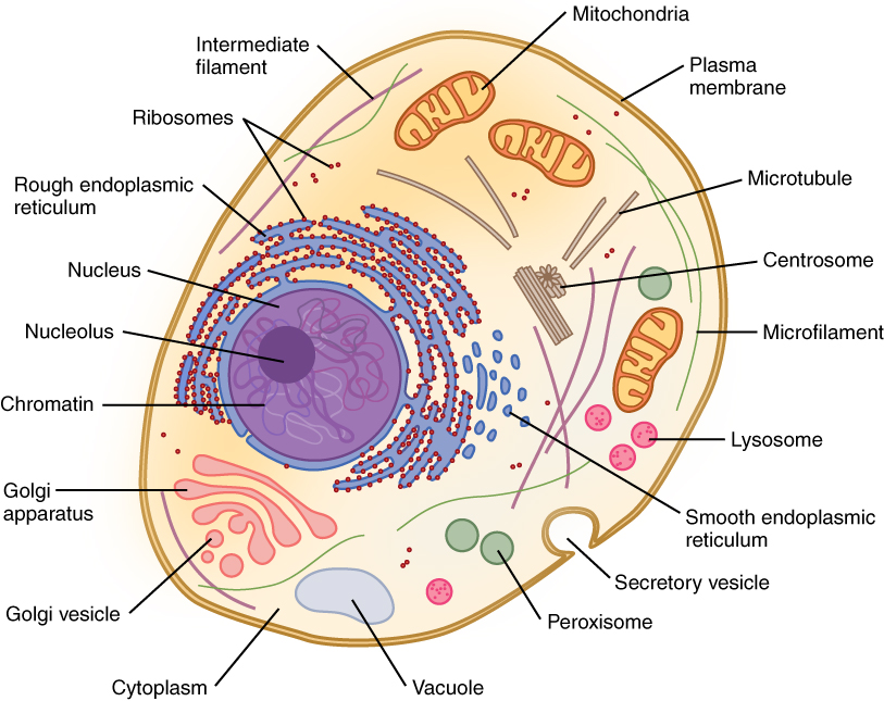
Endoplasmic reticulum (ER)
The Endoplasmic reticulum refers to a tubular membrane system. The rough ER is occupied by ribosomes and facilitates protein biosynthesis of endosomes, transmembrane proteins or secretory granules. The smooth ER is not occupied by ribosomes and has the following diverse functions:
- Stores and regulates calcium ions in the cytopasm of striated muscle cells (here called sarcoplasmic reticulum)
- Synthesis of lipid and steroid hormones
- Detoxification of endogenous and foreign substances within the hepatocytes

Golgi apparatus
The Golgi apparatus is composed of dictyosomes (a stack of four to ten membrane-enveloped, disc-shaped cavities) and has a convex cis-region and a concave trans-region, which face each other. Proteins produced in the Rough ER, reach the cis-Golgi by means of transport vesicles, after which they are then modified and processed (phosphorylation, sulfation, glycosylation) within the Golgi apparatus and sorted with regards to their destination.
At the trans-site, the packaging in secretory granules or vesicles takes place. A retrograde transport (trans to cis) can be detected for enzymes that are required in the ER.

Mitochondria
These “power stations” of the cell supply energy to cells by means of oxidative phosphorylation and are a common topic in exams. Except in mature erythrocytes, mitochondria are found in all cells.
Mitochondria possess two membranes as well as an intermembrane space in between. The smooth outer membrane contains porins, by which molecules <10 kDa can pass through, whereas the inner membrane is folded to a great extent in order to increase the surface area. It restricts the matrix space and carries enzymes of the respiratory chain and the ATP synthesis.
Basically, there are two distinct types of inner membrane due to the folding:
- Crista type: in metabolically active cells such as cardiomyocytes
- Tubule type: in steroid-producing cells
Enzymes of the β-oxidation and those of citric acid cycle are located in the matrix space.
Mitochondria are semi-autonomous, since they have their own circular DNA (mtDNA). As stated in the endosymbiotic theory mitochondria are phylogenetic prokaryotes that are incorporated in eukaryotes in the course of symbiosis.
This hypothesis is further supported by the fact that mitochondria possess 70S ribosomes (50S and 30S subunits) and the involvement of bacterial lipid cardiolipin in the development of the inner membrane.

Lysosomes
An acidic pH (4.5 – 5) as well as a high content of acidic hydrolases, proteases, lipases, esterase enzymes, elastases, collagenases and acidic phosphatases, among other things, are the characteristics of lysosomes.
Their main features are auto- and heterophagy as well as the degradation of endogenous and foreign substances. When the primary lysosome (yet inactive) merges with the substances to be degraded it is referred to as a secondary lysosome.
Peroxisomes

Peroxisomes are mainly located in the liver and kidney and contain the enzymes peroxidase and catalase, as they serve the purpose of degrading fatty acids by means of oxidation. During this process the by-product, hydrogen peroxide, is produced and can lead to cell damage. Therefore, it has to be degraded to water and oxygen by means of a catalase.
Ribosomes
Eukaryotic 80S ribosomes are composed of two subunits (60S and 40S) consisting of one third proteins and two thirds rRNA. They can be found in the cytosol to help in the synthesis of cytoplasmic and nuclear proteins or can be membrane-bound in the rough ER to enable the synthesis of lysosomal proteins as well as to allow the exportation of proteins or membrane proteins.
Centrioles
These cell organelles appear in cylindrical form and are composed of microtubules. One pair of centrioles is perpendicularly arranged to another to form the centrosome. The centrosome is the formation site of microtubules and is also called MTOC (microtubule organizing center).
Cell inclusions
Cell inclusions are byproducts of metabolism, stored nutrients, accumulations of exogenous or endogenous substances that are free within the cytoplasm. This includes glycogen particles, intracellular fat droplets, pigmented cell structures (hemosiderin, lipofuscin, carbon dust) and virus particles.
In the case of certain diseases such as hemochromatosisor glycogen storage disease, cell inclusions are present to a pathological extent.
Components and Functions of the Cytoskeleton
The cytoskeleton is located within the cytoplasm and is responsible for the stabilization, intracellular transport of substances as well as migration (lat.: migrare=hiking) of the cell. This three-dimensional network is generated by microtubules, intermediate filaments and actin filaments. These components are subjected to constant assembly and disassembly, the so-called polymerization and depolymerization.

Actin filaments (F-actin)
They are the smallest components of the cytoskeleton having a diameter of 7 nm and are also called microfilaments.
F-actin is composed of two actin chains, helically wound around each other, that are generated by the polymerization of many globular actin monomers (G-actin). In many cases – but not always – actin filaments are associated to myosin, the actin system’s motor protein. They form the basis of the muscles’ mechanism of filament sliding.
In addition, actin filaments also have stabilizing functions, since they form the basic structure of microvilli or the anchorage point of desmosomes.
Intermediate filaments
With a diameter of 10 nm, they form the passive supporting structure of the cell. The intermediate filaments’ expression varies among tissue types, hence through their means, the origin of a malignant tumor can be determined, for example:
Microtubules
With a 25 nm diameter, they are the largest components of the cytoskeleton and originate from the centrosome (see above). They are composed of α- and β-tubulin dimers and are similar in appearance to a hollow cylinder, one end is charged negatively and the other positively.
Microtubules determine the location of cell organelles within the cell and form a network of direct mass transfer. They are also the basic structures of kinocilia and the spindle apparatus during mitosis and meiosis.
Structure and Function of Cell Contacts
Based on the functions of the three cell types, contacts can be classified into the following categories:
- Communication contacts
- Adhesion contacts
- Barrier contacts as impermeable connections
Communication contacts
They include gap junctions, also called nexus, an absolute favorite in tests.
They consist of transmembrane proteins called connexin. Six connexins form a connexon and two connexons then form a nexus. They enable electrical and metabolic communication between two adjacent cells. A particularly large number of gap junctions are found in the intercalated disc of the myocardium, for example.
Adhesion-/adhesion contacts
They serve as mechanical anchors and consist of three essential components: transmembrane proteins, plaque proteins and the cytoskeleton. Desmosomes can be found between adjacent cells as cell-cell contacts. Hemidesmosomes on the other hand link the cell to the extracellular matrix, creating a cell-matrix contact.
The following table shows an overview of the different types of adhesion contacts, since they are relevant for examination in histology and by part in biochemistry.
Barrier-/closure contacts
They are called tight junctions respectively, zonula occludens, and are developed by the merging of the outer membrane of adjacent cells. Thus, the intracellular space is belt-shaped and encloses this area, so that the paracellular molecule flow is hindered (diffusion barrier). In this area occludin and claudin are important transmembrane proteins.
Junctional complex
This adhesive complex serves as a selective permeability barrier and when viewed from the apical to basal layer consist of: Zonula occludens, zonula adhaerens, macula adhaerens.
Cell Communication
Hormones and transduction
Hormones are chemical messengers that transfer information from one cell to the other. These compounds are produced by the endocrine glands such as those of the pituitary and thyroid.
Overview of important endocrine organs and hormones
Endocrine organs produce hormones in response to signals from the external environment like pain, pressure, heat and light. Hormones can also be produced in response to signals from within the body such as those of hunger.
- Once the hormones are produced, they are secreted in the blood which carries them to cells in other parts of the body where they exert their effect.
- The target cells for these hormones have receptors which enable them to respond to the hormone. These receptors are made up of proteins and are usually located in the plasma membrane which is found on the surface of the cell though some can also be found inside it.
- When the hormone reaches a cell with its receptor, it binds to the receptor and causes a conformational change. This means that the receptor changes its shape and limits its ability to bind to another hormone.
- The conformational change also triggers a series of reactions within the cell which are known as transduction cascades. Depending on the hormone and the specific cell with its receptor, this chain of events can involve the release of enzymes that work together to generate responses.
Effects of hormones on target cells
These responses range from cell division, cell motility and cell death. They can also involve changes in the ion channels that permit or restrict the movement of certain molecules into cells.
Other effects of hormone signal transduction include the absorption of glucose from the blood (insulin), increasing blood pressure and heart rate (adrenaline) and regulation of the menstrual cycle (estrogen and progesterone).
Signal transduction enables the cell to control its response to the hormone as well as to the environment. By having many steps, the effects of the hormone are also amplified.
Signal transduction is therefore very important to organisms that have many cells, since it enables them to coordinate their activities and respond to various signals simultaneously.
Review Questions
The solutions are below the references.
1. Which statement about mitochondria is not true?
- They have their own circular DNA.
- They occur in all cells.
- The matrix space is the site of β-oxidation.
- The bacterial lipid cardiolipin is part of the inner mitochondrial membrane.
- They possess 70S ribosomes.
2. What allocation of tissue type and intermediate filament is wrong?
- neurocytes – neurofilament
- fibrocytes – vimentin
- myocytes – Desmin
- osteocytes – GFAP
- epidermis – cytokeratin
3. What size order of the cytoskeleton components is correct?
- Microtubules < actin filament < intermediate filament
- Actin filament < microtubules < intermediate filament
- Intermediate filament < actin filament < microtubules
- Actin filament < intermediate filament < microtubules
- Intermediate filament < microtubules < actin filament


Comentários
Enviar um comentário