Hypothalamus, Brain Stem, the Circumventricular Organs (CVOs) and the Cerebrospinal Fluid (CSF)
In Lecturio ;) The Hypothalamus is located below the thalamus. The brain stem, consisting of midbrain, pons and medulla oblongata follows, connecting the superior brain structures with the spinal cord. A clear, colorless liquid flows in the brain as well as in the spinal cord – the cerebrospinal fluid. Its functions and clinical relevance will be discussed in this article along with the functions of the different circumventricular organs.
Table of Contents
Image : “Brain” by
VSRao. License: CC0
Adrenal Cortex
Hypothalamus
The Hypothalamus is an almond-sized structure located below the thalamus. It plays a major role in homeostasis of the body forming the bridge between the nervous system and the endocrine system. Anatomically, it is divided into three regions, each consisting of numerous nuclei. The different regions and their respective nuclei along with their functions are as outlined below.
Anterior region of the hypothalamus
The pre-optic area consists of four nuclei namely median pre-optic nucleus, medial preoptic nucleus, ventrolateral pre-optic nucleus and pre-optic periventricular nucleus. This area of the hypothalamus is responsible for thermoregulation and receives sensory signals from the thermoreceptors. It is also thought to influence parenting and sexual behavior as well as the release of gonadotropic hormones. It shows sexual dimorphism.

Image: “Location of the hypothalamus.” by Blausen.com staff. “Blausen gallery 2014”. Wikiversity Journal of Medicine. DOI:10.15347/wjm/2014.010. ISSN 20018762. – Own work.
License: CC BY 3.0
The medial area comprises of the following nuclei:
- The supra-optic nucleus releases vasopressin and oxytocin.
- The paraventricular nucleus causes the release of a thyrotropin-releasing hormone, corticotropin-releasing hormone and somatostatin hormone.
- The anterior hypothalamic nucleus inhibits thyrotropin. It also plays a role in thermoregulation and controls sweating and panting reactions.
- The suprachiasmatic nucleus is responsible for the circadian rhythms.
The lateral area consists of the orexinergic nucleus. The orexin neurons control the feeding behavior as well as the arousal reaction. It also controls visceral functions such as respiration, micturition, blood pressure, in collaboration with other structures of the brain.
Tuberal region of the hypothalamus
The tuberal region is divided into the medial and lateral area. The medial area comprises of the following three nuclei:
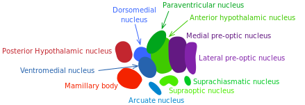
Image: “The hypothalamic nuclei.” by oldblueday – Own work. License: CC BY-SA 4.0
- The dorsomedial hypothalamic nucleus, which controls the blood pressure, heart rate and gastrointestinal motility.
- The ventromedial nucleus controls satiety.
- The arcuate nucleus releases growth hormone-releasing hormone (GHRH). It also controls feeding mechanisms and causes prolactin inhibition via dopamine secretion.
The lateral area of the tuberal region performs similar functions as the lateral area of the anterior region.
Posterior region of the hypothalamus
It is also divided into the medial and lateral area. The medial area consists of:
- The mammillary nuclei, which controls memory.
- The posterior nucleus, which increases blood pressure, causes pupillary dilatation and shivering.
The lateral area comprises of the tuberomammillary nucleus, which controls learning, memory, sleep, arousal, feeding, and energy balance.
Brain Stem

The brain stem is the posterior part of the brain which connects the superior brain structures such as the hypothalamus discussed above to the spinal cord. It consists of the midbrain, pons, and medulla oblongata. The brainstem is responsible for carrying out vital functions such as breathing, arousal, control of heart rate and blood pressure.The figure shows the components of the brain stem.
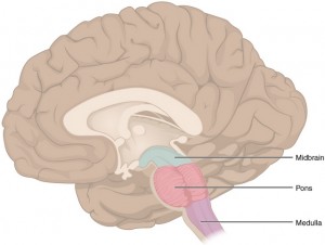
Midbrain
The midbrain is divided into three parts: tectum, tegmentum, and ventral tegmentum. The tectum is made up of a bulge of brain matter that is responsible for auditory and visual reflexes. Tegmentum controls the voluntary movements of the body and is the site origin of cranial nerve III and IV nuclei.
The ventral tegmental area is responsible for reward, cognition, and motivation. Several psychiatric disorders occur due to malfunctioning of this area of the midbrain.
Pons
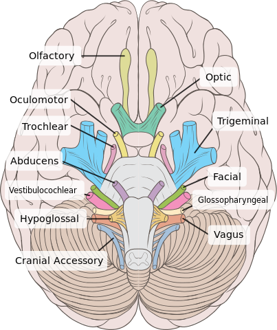
Pons connect the midbrain to the medulla oblongata. It lies in front of the cerebellum. The nuclei of cranial nerves V-VIII originate from the pons.
Pons have their physiological role in controlling hearing and maintaining balance, taste, sensory sensation over the face, secretion of saliva and tear production, eye movements, chewing and facial expressions.
It carries motor signals from the cerebrum to the medulla oblongata and cerebellum and also sensory signals to the thalamus. The figure beside illustrates the site of origin of cranial nerves.
Medulla oblongata
The medulla oblongata is the lowermost part of the brain stem and is connected to the spinal cord. It consists of the cardiovascular center, which controls sympathetic and parasympathetic stimulation of the blood vessels and the heart.
The control of ventilation through chemoreceptors maintains an adequate level of oxygen and carbon dioxide in the blood. The vasomotor center controls blood pressure through sensory signals from the baroreceptor. It also controls vomiting, sneezing, coughing and swallowing reflexes.
Circumventricular Organs (CVOs)
The circumventricular organs are structures of the brain that are located around the third and fourth ventricle. They characterized by an extensive vasculature and lack of blood-brain barrier (BBB). This allows the exchange of substances between the central nervous system (CNS) and the peripheral circulation. The CVOs are broadly classified into two categories: sensory organs and secretory organs.
Sensory organs
Sensory organs have the ability to detect plasma composition. They send the sensed information to higher brain centers so as to get appropriate responses. There are four sensory organs in this category:
- Area postrema senses the presence of the noxious stimulus and triggers vomiting.
- Vascular organ of the lamina terminalis senses the tonocity of the plasma and helps with the maintenance of body fluid homeostasis.
- Subfornical organ has a role in the maintenance of blood pressure and energy balance of the body.
Secretory organs
These organs respond by means of the feedback mechanism and secrete various hormones and other molecules directly into peripheral blood circulation.
- The subcommissural organ (SCO) helps in maintaining the patency of Sylvian duct through secretion of SCO-spondin. It is also hypothesized that it regulates the secretion of aldosterone, detoxification of cerebrospinal fluid (CSF) and osmoregulation.
- Posterior pituitary stores oxytocin and anti-diuretic hormone (ADH) synthesized by the hypothalamus. It plays a role in homeostasis.
- Median eminence is classified as a circumventricular organ, though this is still controversial. Due to a rich network of capillaries, it allows the transport of neurohormones between the CSF and peripheral blood.
- Pineal gland shows a circadian rhythm in its activity. It secretes melatonin under the command of supra-chiasmatic nuclei (SCN). The secretion of melatonin is suppressed during the day or when there is light. The pineal gland is also thought to play a role in sexual development as melatonin has been found in pre-ovulatory follicles, semen, breast milk and amniotic fluid.
Cerebrospinal Fluid (CSF)
Cerebrospinal fluid is a clear, colorless liquid that flows in the brain and the spinal cord. It is produced by the choroid plexus in the brain and flows in:
- Subarachnoid space between the arachnoid and pia mater
- Ventricular system
- Spinal cord
Composition of CSF
The pH of CSF is around 7.33 with an osmolarity of 295 mOsm/l. It is mainly composed of:
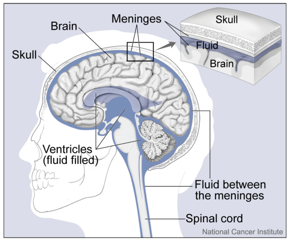
Image: “Distribution of cerebrospinal fluid in the brain and spinal cord.” by Alan Hoofring – https://visualsonline.cancer.gov/details.cfm?imageid=4279. License: Public Domain
- Water (99 %)
- Protein (35 mg/dL)
- Glucose (60 mg/dL)
- Sodium (138 meq/L)
- Potassium (2.8 meq/L)
- Calcium (2.1 meq/L)
- Magnesium (0.3 meq/L)
- Chloride (119 meq/L)
Functions of CSF
When the brain is suspended in CSF, its mass is transformed from 1,400 g to 25 g. This is the neutral buoyancy, which allows the brain to perform its functions without compressing the lower parts. The blood supply to different areas of the brain is not affected by this light weight of the brain.
It protects the structure of the brain from coming in direct contact with the skull. However, in severe head injuries, the CSF moves to the opposite side of impact causing brain damage, which may lead to death.
The CSF is drained into the peripheral vasculature. This prevents the accumulation of harmful substances in the brain.
The CSF fluid may decrease in volume in certain areas of the brain to relieve the pressure effects thereby improving the blood supply to that part of the brain.
Clinical correlation of CSF
CSF analysis is an investigative technique performed to know the constituents of CSF. It may give different results for different clinical conditions and therefore helps in making the right clinical diagnosis. The procedure performed to obtain CSF is known as a lumbar puncture.
The abnormal accumulation of extra CSF in the ventricles of the brain is called hydrocephalus. It leads to increased intracranial pressure. If this occurs during the mental development of the fetus it leads to an enlarged cranium and the condition is termed as congenital hydrocephalus. It results in mental disability and convulsions and therefore needs prompt intervention. If left untreated, it can be fatal.
Baricity is the density of a certain drug compared to the density of the cerebrospinal fluid. It is of importance in anesthesiology where the duration of anesthesia depends upon the rate of spreading of a certain drug in the intrathecal space and the position of the patient. For example, when using a drug of higher baricity than CSF for spinal anesthesia then the patient is position with the lower limbs at a lower level than the body. When the anesthetic effect is desired more proximally, the position can be altered to maximize the flow and distribution of the anesthetic agent albeit theoretically.
The figure above shows the distribution of CSF in the brain areas. The flow begins with CSF production within the cerebral ventricular system, mainly by the choroid plexus at rate of 20ml/hr. Each of the four ventricles contains choroid plexus tissue which is made up of villous folds lined by epithelium and a central core of highly vascularized connective tissue. Here, active secretion and diffusion produce CSF. From the two lateral ventricles, fluid drains through the foramen of Monro on each side into a single midline third ventricle.
The third ventricle connects by the aqueduct of Sylvius to a midline fourth ventricle that has three exit openings, paired lateral foramina of Luschka, and a midline foramen of Magendie. These openings lead to a system of interconnecting and focally enlarged areas of subarachnoid spaces referred to as cisterns which allow for free flow of CSF. Fluid in the basal cisterns, tentorium, and subarachnoid space flows over the cerebral convexities to the sagittal sinus, from where absorption into the systemic circulation takes place.
Most CSF absorption takes place across the arachnoid villi into the venous channels of the sagittal sinus, but fluid is also absorbed across the ependymal lining of the ventricular system and from the spinal subarachnoid space. The process of CSF formation continues when the intracranial pressure rises, unless extremely high levels are reached.Thus, there must be some absorption of fluid to accommodate the volume of CSF being formed each day or more room for expansion must be available or must be created


I enjoyed this blog post. It is inspiring and informative.
ResponderEliminarhttps://blog.mindvalley.com/brain-stem/