Histology of the Kidney
Table of Contents
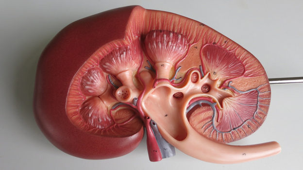
The Kidney
These are two retroperitoneal organs that weigh about 150 grams each. They are lined and protected by a fibrous capsule. Thefunctions of this organ are ultrafiltration and excretion of metabolic waste products, electrolyte and water balance, secretion of hormones and acid base balance among others.
The Nephron – Functional Unit of the Kidney
The smallest functional unit of the kidney which filters urine from the blood, is the nephron. A kidney contains about 1.4 x 10⁶ nephrons. Its structure can be classified into two groups for better understanding. The Glomerulus and Bowman’s capsule form the renal corpuscle which passes into the renal canaliculi, which consists of proximal, intermediate and distal tubules.
The renal corpuscle
Any unfiltered blood passes through the arteriole/afferent vessel into a bundle of capillaries (glomerulus) and leaves after filtration through the efferent vessel at the same spot. These capillaries are fixed to the mesangium and extend into the Bowman’s capsule. Glomerulus and Bowman’s capsules form the filtration barrier.
The Bowman’s capsule can be imagined as a bubble in which the glomerulus is pressed into – it has a visceral layer which is in contact with the glomerulus, and a parietal layer which forms the wall of the “ball“. At the opposite of the entry and exit of the capillaries (vascular pole), a tubule leaves the capsular space (urinary pole). It drains the primary urine from the capsular space.
The filtration barrier
The filtrate has to overcome three barriers to reach the capsular space: the endothelium, the glomerular basement membrane and the slit membrane of the podocytes.
- The endothelium is fenestrated (window size 70 – 100 nm) without the diaphragm. At its inner side, a negatively charged glycocalyx impedes filtration of bigger, negative molecules.
- The glomerular basement membrane consists of lamina rara interna, lamina densa and lamina rara externa. The net out of collagen IV of the lamina densa stabilizes the structures and has to sustain the blood pressure within the capillaries. In both laminae rarae, there are a large number of molecules and proteins for negative charging of the basement membrane e.g.
- The slit membrane is formed by branches of the podocytes. They are the visceral layer of the Bowman’s capsule. The foot processes of the podocytes are interlocking. Little interstices are spanned by the slit diaphragm which is formed by the membranous protein nephrin. Molecules that have a diameter of up to 4.4nm fit through the filtration pores of the slit diaphragm.

The tubule system of the nephron
The tubule has the task of converting the primary urine into final urine. For this to happen, it net resorbs about 99 % of the filtrated water, glucose as well as electrolytes and much more. The epithelium is connected by tight junctions in every part; it has a distinct basal labyrinth(folding of the basement membrane) and many mitochondria (high energy consumption possible).
This can be seen as dark basal striation of the cells under a light microscope. Furthermore, the segments differ in terms of structure and function. In the following synoptic table, the segments of the tubule starting from the urinary pole of the renal corpuscle, and their main function are listed. Subsequently, the specific epithelia are described in detail.
The proximal tubule
The cubic epithelial cells are well interlocked and have a distinct basal labyrinth as well as a high brush border. The basolateral Na-K-ATPase is the driving force for reabsorption of amino acids, sodium and glucose. Water osmotically passes through the zonulae occludentes (tight junctions) of the epithelium.
The intermediate tubule
The cells of the intermediate tubule are rather flat, the lumen of the tubule is the smallest here (12 – 15 µm). The nucleuses often curve into the lumen, and the cytoplasm is thicker than the cytoplasm inside the blood capillaries. In the descending part of the loop of Henle, the epithelium is permeable to water while in the ascending part, it is impermeable. This way, it contributes to the concentration of the final urine.
The distal tubule
The epithelium has a cubic form with round nucleus again and has a distinct basal labyrinth as well as single microvilli inside the tubule lumen. The tight junctions are particularly distinct here and the epithelium is absolutely impermeable to water.
The Collecting Duct – Fine Adjustment of the Final Urine
The collecting duct does not relate to nephron either biologically or functionally. A collecting duct receives influx from about 11 nephrons in the cortex. From here, it proceeds through the outer and inner medullary collecting ducts along the medullary rays to the renal medulla and papilla.
On their way, collecting ducts repeatedly merge and the lumen widens up to 200 µm. The epithelium changes from a cubic to a prismatic shape. It has a rarely distinct basal labyrinth. Two types of cells can be found:
- Chief cells:
- Microvilli, some kinocilia; appear lighter under a light microscope
- Place of the ADH-dependent integration of aquaporins and, therefore, reabsorption of water: in the collecting duct, about 20 – 30 % of the H2O reabsorption takes place (70 – 80 % in the proximal tubule). Only here, can the reabsorption be matched with the physiological needs of the body by ADH secretion.
- Regulation of the sodium resorption and potassium secretion by aldosterone
- Switch cells:
- Rich in mitochondria; appear darker under a light microscope
- Type-A-switch cells: secretion of protons, resorption of bicarbonate
- Type-B-switch cells: secretion of bicarbonate in alkaline metabolism
The Juxtaglomerular ApparatusThe distal straight tubule of every nephron directly passes the afferent arteriole of its glomerulus at one spot. The glomerular filtration is regulated at this junction. The epithelium of the tubule (columnar and dark here = macula densa), the juxtaglomerular smooth muscle cells as well as the extraglomerular mesangium cells are part of this.The distal epithelium imparts the actual ion concentration of the urine and baroreceptors of the juxtaglomerular cells record the blood pressure in the afferent vessel. Both factors can cause a release of renin from the juxtaglomerular cells.Topography of the NephronAll nephrons and the corresponding collecting ducts are arranged in the same pattern. The transitions between different tubular parts of different nephrons lie nearly on a par. Due to this parallel structure of all nephrons, the histological transitions between the cortex and medulla are macroscopically visible.The nephrons and convoluted parts of the proximal and distal tubule lie in the cortex. The transition to the straight part marks the cortex-medulla-line. Collecting ducts and loops of Henle that belong to the superior nephrons can be seen as collimated medullary streaks in the cortex. These functionally belong to the medulla but reach into the cortex. The other collecting ducts and loops of Henle can be found within the medulla. The Interstitium of the KidneyIt is made up of extravascular intertubular spaces or the renal parenchyma, cellular elements, and extracellular substances.Only a small connective tissue can be found inside the peritubular interstitium, however, there are many macrophages and fibroblasts. The fibroblasts are responsible for erythropoietin production. Erythropoietin stimulates the formation of red blood cells within the bone marrow. Furthermore, many interstitial cells produce prostaglandins.Review QuestionsThe answers can be found below the references.1. In which part of the tubule is the most amount of water reabsorbed?
- Proximal straight tubule
- Proximal convoluted tubule
- Collecting duct
- Loop of Henle
- Distal convoluted tubule
2. Which of the following attributes does not apply to the epithelium of renal tubules?- Distinct basal labyrinth
- Brush border
- Fat vacuoles
- Tight junctions
- Rich of mitochondria
3. Which assertion is correct?- The afferent vessel has a fenestrated endothelium with diaphragms.
- Nephrin is an important protein of the basement membrane.
- There are no proteins in the lamina rara of the basement membrane.
- Positive molecules are more filtrated than negative molecules.
- Molecules with a diameter of up to 10nm are filtrated.


Comentários
Enviar um comentário