12 Cranial Nerves — Functions and Mnemonics
Table of Contents
- Classification of Cranial Nerves
- List of Cranial Nerves
- Cranial nerve I: Olfactory Nerve
- Cranial nerve II: Optic Nerve
- Cranial Nerve III: Oculomotor Nerve
- Cranial Nerve IV: Trochlear Nerve
- Cranial Nerve V: Trigeminal Nerve
- Cranial Nerve VI: Abducent Nerve
- Cranial Nerve VII: Facial Nerve
- Cranial Nerve VIII: Vestibulocochlear Nerve
- Cranial Nerve IX: Glossopharyngeal Nerve
- Cranial Nerve X: Vagus Nerve
- Cranial Nerve XI: Accessory Nerve
- Cranial Nerve XII: Hypoglossal Nerve
- Overview of the 12 cranial nerves
- Review Questions
- References
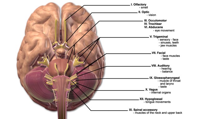
Classification of Cranial Nerves
Every cranial nerve (CN) is assigned a Roman numeral as a name. The numbering is based on the order in which they emerge from the brain, from ventral to dorsal. The name indicates the function or the course.
List of Cranial Nerves
- I Olfactory
- II Optic
- III Oculomotor
- IV Trochlear
- V Trigeminal
- VI Abducens
- VII Facial
- VIII Vestibulocochlear
- IX Glossopharyngeal
- X Vagus
- XI Accessory
- XII Hypoglossal
How do the cranial nerves differ in terms of fibers?
The 12 pairs of cranial nerves originate from the nose (CN I), the eyes (CN II), the inner ear (CN VIII), the brainstem (CN III-XII) and the spinal cord (part of XI).
Cranial nerves have sensory (afferent) and motor (efferent) functions. Only sensory axons contain two cranial nerves (CN I and CN II), which are sensory functions, but fiber tracts. The olfactory nerve emerges from eversions of the cerebrum and the optic nerve emerges from the diencephalon.
All the other cranial nerves originate from the brainstem nuclei (the hypoglossal nerve is located at the border of the spinal cord) and include sensory axons, as well as motor axons. The oculomotor nerve (III) and the trochlear nerve (IV) emerge from the mesencephalon, the cranial nerves V to XII originate from the pons (bridge) and the medulla oblongata (extended spinal cord).
The cranial nerves III, IV, VI, XI and XII are mainly motor and provide innervation of the skeletal muscles. However, the cranial nerves I and II mentioned before, and the vestibulocochlear nerve, are purely sensory, but there are also cranial nerves (CN V, VII, IX, X) that are both sensory and motor.
The oculomotor nerve, the facial nerve, the glossopharyngeal nerve, and the vagus nerve also include somatic and autonomic axons. The somatic part ensures the supply of the skeletal muscles and the autonomic part belonging to the parasympathetic nervous system innervates the glands, the smooth muscles, and the cardiac muscle.

Get a short overview of the cranial nerves I—VI
Cranial nerve I: Olfactory Nerve
Quality and Course

The olfactory nerve is part of the olfactory pathway and is a purely sensory nerve The olfactory mucosa, with its olfactory cells, is located in the superior nasal meatus (meatus nasi superius).
The olfactory cells are nerve cells of which the unmyelinated axons are bundled and emerge through the openings of the cribriform plate (lamina cribrosa, part of the ethmoid bone) and the dura mater located on top into the anterior cranial fossa. The approximately 40 bundles make up the right and the left olfactory nerve.
The olfactory nerve is the shortest nerve that does not travel via brainstem. In olfactory nerves end in the brain in two paired masses of grey matter, the olfactory bulb, where they are switched to the second neuron. The axon terminals (synaptic knobs) of the olfactory receptors (olfactory cells) compose the next neurons on the olfactory pathway in the respective bulbus synapses with dendrites and soma.
These axons emerging from the neurons of the olfactory bulb are located in the olfactory tract, which leads to the olfactory cortex (rhinencephalon/smell brain), where the olfactory sensation is perceived and then emotionally linked.
Function of the olfactory nerve
- Smelling and more advanced aspects of taste
Cranial nerve II: Optic Nerve
Quality and Course
The optic nerve is the visual nerve (=optikos= vision) and it is purely sensory in function. The optic nerve is formed by the convergence of axons from the retinal ganglion cells. These cells, in turn, receive impulses from the photoreceptors of the eye. For the most part, it is encased in the three meninges. From the retina of the eye, the visual impulses are transmitted to the diencephalon via the optic nerve. Fibers pass from the visual center of the thalamus to the occipital visual cortex, where the image of the event is created.

However, both the optic nerves do not run separately to their ipsilateral part of the thalamus. In the part of the sphenoid bone, the sella turcica (Turkish Chair) they join into the optic decussation (chiasma opticum). There, the fibers of both the optic nerves mix until new fiber tracts form and continue to the diencephalon. As of the chiasma, the nerve bundles are not called nerves any longer, but optic tract.
The transmission of the field of vision leads to the mixture of the fibers. In the optic tract, the part of the optic nerve transmitting the lateral (temporary) field of vision proceeds on the same side.In the optic decussation, the medial parts of the field of vision are switched to the contralateral side and cross to the opposite side.
Function of the Optic Nerve
- It functions to transmit visual information from the retina to the vision centers of the brain through electrical impulses.
Cranial Nerve III: Oculomotor Nerve
Quality and Course
The oculomotor nerve (oculus= eye) is a mixed cranial nerve, which is mainly motor, while the motor nucleus is located in the ventral mesencephalon. It innervates most of the outer (extraocular) eye muscles and passes from the midbrain (mesencephalon) to the bony eye socket (orbit).
It extends ventrally and divides into a superior and an inferior branch. The rectus superior muscle (one of the outer eye muscles) and the levator palpebrae superioris muscle (muscle of the upper eyelid) are innervated by the axons of superior branch of the oculomotor nerve. The rectus medialis muscle, the rectus inferior muscle and the obliquus inferior muscle, which are all outer eye muscles, are supplied by the axons of the inferior branch.
Function of the Motor Axons
- Movement of the upper eyelid and the eye
The oculomotor nerve serves the voluntary motor function, but it also contains parasympathetic fibers.
The inferior branch serves the parasympathetic innervation of the inner eye muscles and contains the ciliary muscle and the iris sphincter muscle. Parasympathetic impulses, reaching the ciliary ganglion, originate from the oculomotor nucleus in the mesencephalon, which is the “circuit center” of the autonomous nerve system.
Function of the Parasympathetic Axons
Parasympathetic axons extend from the ciliary ganglion to the ciliary muscle, which is responsible for the
- Accommodation (adaption to the distance of an object).
Further parasympathetic fibers serve:
- The stimulation of the iris sphincter muscle (constriction of the pupil in bright light) and
- The stimulation of the iris dilator muscle (dilatation of the pupil).
Function of the Sensory Axons
- Proprioception (part of self-perception)
The sensory part consists of afferent neurons of the proprioceptors of the outer eye muscles towards the mesencephalon. These axons bring together information about non-visual perception of body movement and position in space, as well as the location and position of single body parts to each other. It controls the muscles that allow for visual tracking and fixation by the eye. Visual tracking is the ability to follow an object as it moves across the field of vision.
Cranial Nerve IV: Trochlear Nerve
Quality and Course

Image: “Trochlear Nerve” by Btarski. License: CC BY-SA 3.0
The trochlear nerve is the smallest of the 12 cranial nerves and the only nerve exiting the dorsal aspect of the brainstem with mostly motor axons. The trochlear nerve arises from the trochlear nucleus of the brain, emerging from the posterior aspect of the midbrain. It is the only cranial nerve to exit from the posterior midbrain.
Beneath the tectum of mesencephalon (quadrigeminal plate), the nerve exits the midbrain (mesencephalon) and, as the olfactory nerve, it passes through the wall of the cavernous sinus.
Function of the trochlear nerve
Somatomotor functions:
- Eye movement, innervation of the obliquus superior muscle
Sensory functions:
- Proprioception
Cranial Nerve V: Trigeminal Nerve
Quality and Course
The trigeminal nerve is a mixed cranial nerve with mainly sensory parts and it is regarded as the largest cranial nerve. It runs from the pons to the petrous portion (Pars petrosa) of the temporal bone (Os temporale), where it converges the trigeminal ganglion. As the name suggests, the trigeminal nerve has three major branches consisting of nerve cell clusters.
- Ophthalmic nerve
- Maxillary nerve
- Mandibular nerve
The ophthalmic nerve passes through the superior orbital fissure and is the smallest branch. The maxillary nerve is located between the ophthalmic nerve and the mandibular nerve. It is medium-sized and passes through the foramen rotundum. The mandibular nerve passes through the oval foramen and is the largest of the three trigeminal branches.
Function of the trigeminal nerve
The transmission of impulses for touch, pain, temperature, proprioception and the somatomotor function with mandibular movement, are the sensory functions of the trigeminal nerve.
The ophthalmic nerve (V1) has mainly sensory fibers and has the following function:
- Sensory innervation of the skin of the forehead, the eyes, the nose, and the nasal mucosa
- Innervation of the iris, the cornea, the conjunctiva, and the lacrimal gland
- Supply of the skin of the forehead and checking for pressure pain at the exit point of the supraorbital foramen
The maxillary nerve (V2) innervates the facial skin with sensory fibers and a parasympathetic part and has the following tasks:
- Supply of the lower eyelid, its conjunctiva, and the upper lip
- Supply of teeth (upper molar, incisor, and canine), nasal cavity gums of the upper jaw and the palate mucosa (incl. uvula) and tonsils
- Supply of the lacrimal gland and nasal glands by the parasympathetic part
The mandibular nerve contains besides the sensory fibers also motor fibers of the trigeminal nerve. It has the following tasks:
- Innervation of all the muscles of mastication (e.g. masseter muscle) and the suprahyoid muscles by motor fibers
- Supply of the teeth (lower molar, incisor and canine teeth), the gums of the lower jaw, the buccal mucosa, the dorsum of the tongue, as well as the external acoustic meatus, including the eardrum, by sensory fibers
Cranial Nerve VI: Abducent Nerve
Quality and Course
The abducent nerve arises from the abducens nucleus of the pons (bridge) and is one of the mixed cranial nerves with mainly motor fibers. From the nucleus, the axons emerge to the rectus lateralis muscle (outer eye muscle), pass through the superior orbital fissure and end in the orbit.
Function of the abducent nerve
Somatomotor functions:
- Eye movement, abduction of the eyes
Sensory functions:
- Proprioception
Get a short overview of the cranial nerves VII-XII
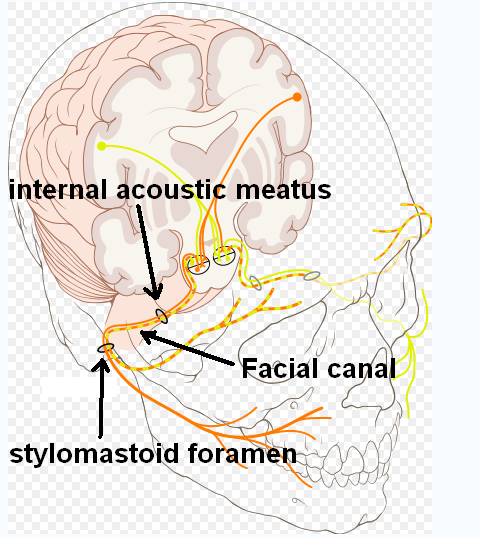
Cranial Nerve VII: Facial Nerve
Quality and Course
The facial nerve is a mixed cranial nerve with motor, sensory and parasympathetic parts. The sensory axons extend from the taste buds of the anterior two-thirds of the tongue through the geniculate ganglion, to the pons (bridge). Somatomotor neurons, on the other hand, originate from a nucleus of the pons and pass through the temporal bone.
The axons of the parasympathetic neurons of the facial nerve end in the two parasympathetic ganglions (pterygopalatine ganglion and submandibular ganglion).

Function of the facial nerve
The facial nerve has mainly motor functions. However, a part of its fibers, which is called an intermediate nerve, serves the sensory functions.
Motor functions:
- Innervation of the entire mimic facial musculature (platysma and muscles of the auricle)
- Eyelid movement and closing of the eye by the orbicularis oculi muscle
- Movement and closing of the mouth by the orbicularis oris muscle
- Precise adjustment of the auditory ossicles by the stapedius muscle
- Movement of the mandible by mentalis muscle
Sensory and parasympathetic functions:
- Sense of taste in the anterior two-thirds of the tongue
- Innervation of the three large salivary glands, the lacrimal glands, and the nasal glands
- External acoustic meatus
Cranial Nerve VIII: Vestibulocochlear Nerve
Quality and Course
The vestibulocochlear nerve is a mainly sensory nerve that has its nucleus in the pons and passes together with the facial nerve to the internal acoustic meatus. There, it splits into two divisions:
- Vestibular nerve
- Cochlear nerve
In the vestibular nerve the sensory fibers of the semi-circular canal, the saccule and the utricle of the inner ear, pass to the vestibular ganglion ending in the vestibular nuclei of the pons.
Axons originating in motor neurons project from the pons to the hair cells of the semi-circular canal, the saccule and the utricle.
The sensory fibers, located in the cochlear ganglion and passing to the nuclei of the medulla oblongata, originate from the cochlear nerve in the organ of corti (spiral organ) located in the cochlea of the inner ear.
Axons of motor neurons extend from the pons to the hair cells of the spiral organ of corti.
Function of the vestibulocochlear nerve
Vestibular nerve:
- Regulation of the hair cells of the organ of corti for the adjustment of sensitivity (spatial position)
- Transmission of impulses for the sense of balance therefore maintains equilibrium.
Cochlear nerve:
- Regulation of the hair cells of the organ of corti for the adjustment of sensitivity (regarding sound waves)
- Transmission of impulses for hearing

Cranial Nerve IX: Glossopharyngeal Nerve
Quality and Course
The glossopharyngeal nerve is a mixed cranial nerve with motor, sensory and parasympathetic fibers, which exits the medulla oblongata behind the olivary bodies, together with the vagus nerve and the accessory nerve.
In the area of the posterior cranial fossa, the sensory fibers exit the cranial cavity; pass between the internal carotid artery and the internal jugular vein, until they laterally reach the root of the tongue. The motor fibers pass through the jugular foramen from the nuclei of the medulla.
Function of the glossopharyngeal nerve
Motor functions:
- Innervation of the palate muscles and the muscles of the pharynx by the vagus nerve
- Dilatation of the pharynx during swallowing and speaking
Sensory functions:
- Innervation of the mucosa of the middle ear, the mastoid, and the eardrum
- Supply of the velum, including the palatine tonsil and the posterior third of the tongue
- Taste and somatic perception (touch, pain, and temperature) of the posterior third of the tongue
- Proprioception in the swallowing musculature
- Blood pressure regulation
- Regulation of the oxygen and carbon dioxide content of the blood for the control of ventilation
Parasympathetic functions:
- Stimulation of salivation
- Carotid glomus – contains chemoreceptors responsible for the oxygen content, as well as pressure receptors that are important for the regulation of blood pressure
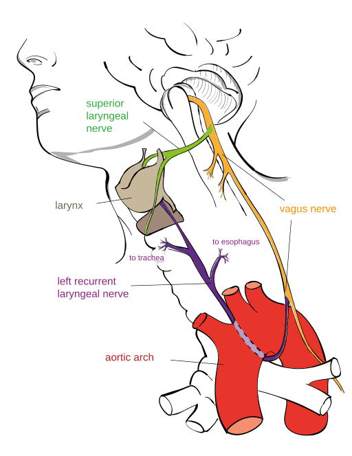
Cranial Nerve X: Vagus Nerve
Quality and Course

Image: “Drawing of the Left Recurrent Laryngeal Nerve” by Jkwchui. License: CC BY-SA 3.0
The vagus nerve is the parasympathetic main nerve, which is responsible for large parts of the organismand which innervates almost all the organs of the thorax and the abdomen. The vagus nerve has the longest course of all cranial nerves.
After exiting the medulla oblongata, the vagus nerve passes through the skull base. Near the carotid artery, it passes to the intrapleural space in the neck, where it gives off branches to the base of the heart (to the atriums) and the hilum of the lung.
Sensory parts of the vagus nerve originate from the skin of the outer ear. They can cause a cough and nausea if they receive a certain stimulus (cotton bud). A few sensory parts originate in the epiglottis and the pharynx; proprioceptors originate in the muscles of neck and pharynx and in chemoreceptors in the carotid glomus near the aortic arch. Further branches pass to the recurrent laryngeal nerve, which passes back to the laryngeal cartilage to supply the vocal folds. In addition, there are axons of viscerosensory receptors of thoracic and abdominal organs.
Additionally, parasympathetic fibers extend from the nuclei of the medulla to the lung and the heart. The pressure receptors at the carotid glomus contain fibers of the vagus nerve that are necessary for the blood regulation. The vagus gives rise to a network of branches around the esophagus to enter the abdomen at the oesophageal hiatus.
The glands of the gastrointestinal tract, the smooth musculature of the respiratory tract, the stomach, the esophagus, the gallbladder, the small intestine and most parts of the large intestine are supplied by parasympathetic axons.
The skeleton muscles of the inner and outer neck are innervated by somatomotor neurons.
Function of the vagus nerve
Sensory functions:
- Taste and sensation (touch, pain, temperature etc.) in the epiglottis and the pharynx
- Blood pressure regulation
- Regulation of the oxygen and carbon dioxide content in the blood for the control of ventilation
- Sensation in visceral, thoracic and abdominal organs
Somatomotor functions:
- Swallowing
- Coughing
- Voice production
Parasympathetic functions:
- Contraction and relaxation of the smooth musculature of the gastrointestinal tract
- Reduction of the heart rate
- Secretion of digestive juices
Cranial Nerve XI: Accessory Nerve
Quality and Course
The accessory nerve is the main motor, mixed cranial nerve, which emerges from the brainstem and the spinal cord.
Cranial root:
The cranial root arises from the nuclei of the medulla oblongata. It passes through the jugular foramen and supplies the musculature of the pharynx, the larynx, and the velum to enable the swallowing process.
Spinal root:
The spinal root contains mixed, but mainly motor axons. The motor axons pass through the foramen magnum and exit the jugular foramen together with the cranial fibers. Motor impulses are transmitted via the spinal root to the sternocleidomastoid muscle and the trapezius muscle, which control the movement of the head.
Sensory axons originate from the proprioceptors of the muscles. They supply the motor neurons and end in the medulla oblongata.

Function of the Accessory Nerve
Sensory functions:
- Proprioception
Somatomotor functions:
- Innervation of the sternocleidomastoid muscle and the trapezius muscle to control the movement of head and shoulders
- Swallowing movement
Cranial Nerve XII: Hypoglossal Nerve
Quality and Course
The hypoglossal nerve is a mixed cranial nerve with mainly motor functions.
The somatomotor axons passing through the hypoglossal canal arise from a nucleus of the medulla oblongata. However, the sensory part consists of axons of proprioceptors of the tongue muscles and ends in the medulla oblongata.
Function of the hypoglossal nerve
Somatomotor functions:
- Innervation of tongue muscles
- Transmission of impulses for speaking and swallowing
Sensory functions:
- Proprioception
Overview of the 12 cranial nerves
Review Questions
The solutions can be found below the reference list.
1. Which mixed cranial nerve, with mainly sensory parts, is regarded as the largest cranial nerve?
- N. vagus
- N. trigeminus
- N. vestibulocochlearis
- N. facialis
- N. trochlearis
2. Which cranial nerves are purely sensory?
- N. olfactorius, N. opticus, and N. vagus
- N. vagus, N. oculomotorius and N. opticus
- N. olfactorius, N. oculomotorius, and N. accessorius
- N. olfactorius, N. opticus, and N. vestibulocochlearis
- N. vagus, N. accessorius and N. vestibulocochlearis
3. Which nerves are not nerves in the proper sense, but fiber tracts and part of the brain?
- N. olfactorius and N. opticus
- N. vagus and N. oculomotorius
- N. olfactorius and N. oculomotorius
- N. opticus and N. oculomotorius
- N. vagus and N. accessorius


Comentários
Enviar um comentário