Facts about Pregnancy: Confirming and Dating Pregnancy, Establishing Gestational Age, First Trimester Bleeding and Fetal Alcohol Syndrome (FAS)
Table of Contents
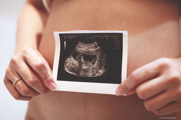
Confirming and Dating Pregnancy and Establishing Gestational Age
After successful conception, the fertilized ovum is implanted into the endometrium where the placenta forms. It is responsible for the production of the hormone beta-HCG which is typical for pregnancies. The increased amount of this hormone is used for the detection of pregnancies. It is considered as a reliable proof of pregnancy. The most reliable proof, though, is a sonographic examination.
Less reliable signs of pregnancy are first movements of the child, palpation of the uterus and first subjective signs of pregnancy. Among these signs is the growth of the mammary glands with a feeling of tightness and morning sickness (emesis).
During the first examinations, the expected date of birth is supposed to be calculated. However, the results of the sonographic examination are not very helpful yet, because the amniotic sac is too small and the crown-rump length cannot be measured yet. Therefore, the first day of the last menstruation is used to calculate the date. The following formula is called Naegele’s rule.
This rule is used for a normal cycle length (28 days). If the menstrual cycle deviates, days should be subtracted or added (extension of Naegele’s rule).
Prenatal Care
Preventive examinations should take place every 4 weeks in the first 4 months. In the following 3 months every 3 weeks, in the following 2 months every 2 weeks and in the last month once per week.
During every examination, the blood pressure is controlled in order to preclude hypertension. The weight, the urine, and the haemogram are examined regularly.
As around 5% of all pregnant women develop gestational diabetes for the first time, the blood glucose is tested routinely.
Ultrasound examinations take place in the 10th, 20th and 30th week of pregnancy. Several parameters are controlled. Control of vitality and detection of malformations, as well as malpresentation of the fetus are essential. In advanced pregnancy, malpresentation of the fetus is particularly important. Moreover, the placenta and the quantity of amniotic fluid are evaluated.
The bimanual gynecological examination is also important for prenatal care. It is carried out until the 16th week of pregnancy. After this, the so-called Leopold’s maneuvers are used. These serve in order to determine the fundal height, the position and the size of the child and the quantity of amniotic fluid.
The first maneuver is used to determine the position of the child. Both hands of the examiner are placed close to the fundus. The fundal height can be estimated with this as well. Navel, xyphoid and costal arches are used for orientation.
The second maneuver is carried out by placing the hands on both sides of the uterus. While doing this, the examiner can estimate the position of the back.
While carrying out the third maneuver, the right hand is placed at the pelvis. Thumb and fingers are used to identify the child’s head and other body parts. Through slightly sudden movements (ballottement) the flexibility of the head can be tested. If there is no flexibility, the child’s head already entered the pelvis.
The fourth maneuver is important to determine the relation between the pelvic and the preceding part. The examiner faces the woman’s feet. Both hands are placed on the ulnar side in direction to the pubic bone.
In order to evaluate the cervix, vaginal examinations are carried out during the preventive appointments. During these appointments, it is important to estimate the width of the uterine orifice, the length of the cervix, the level and the consistency of the cervix.
Control of the fetal vitality is also an important part of the examination. This is assessed using ultrasound at the beginning of the pregnancy. The first heartbeat can be identified as of the 5th week of pregnancy. Movements of the child are also signs of vitality. Typically, these can be determined by CTG (= Cardiotocography).
First Trimester Bleeding
Bleedings during pregnancy can have different reasons. They are categorized according to the stages.
In the first half of the pregnancy reasons for bleeding can be abortion, nidation bleedings, trophoblastic tumors, ectopic pregnancy or cervical carcinoma.
If bleedings occur in the second half of the pregnancy, a placenta previa, a premature placenta abruption, a uterine rupture, a placental edge bleeding or a bloody show while feared delivery should be differentially diagnosed.
During the birth, bleedings can occur as well. Reasons for this can be the uterine rupture, the bloody show, the premature placenta abruption or the insertio velamentosa. The insertion velamentosa is characterized by a strong bleeding after rupturing of the membranes and is a life-threatening danger to the child.
In the case of emergency, a shock prevention, which would include a volume substitution, would be carried out. In a clinic, a surgical stopping of the bleeding would be attempted. If none of the measures are successful, a hysterectomy would be considered.
Fetal Alcohol Syndrome (FAS)
Etiology and epidemiology of the fetal alcohol syndrome
In all stages of pregnancy, alcohol is harmful to the embryo and the fetus. With a prevalence of 1:300 a fetal alcohol syndrome occurs more often than innate malformations such as the Down’s syndrome.
However, alcohol effects, which affect the most sensitive human organ, are more frequent than fetal alcohol syndrome. The effects are characterized by, inter alia, brain achievement weakness, behavioral changes, and behavioral problems.
Alcohol and its metabolites harm the body’s cells, especially tissues with a high regenerative capacity. The substance can be compared to a mitotic toxin. Consequently, the growth of the child is restricted and results into a hypoplasia or a hypotrophy.
An important aspect for the cumulative occurrence of alcohol abuse is the social class. Studies proved that socially disadvantaged women give birth to children with growth restriction more frequently. In middle class’ women this phenomenon occurred only rarely.
Affected children are usually smaller and have a low body weight. In this context, the reduced weight can be traced back to muscle hypertrophy.
Typical characteristics are craniofacial changes of the children. In various tissues the hypertrophy expresses itself particularly strong: frequently, the lower jaw is shifted back (maxillary hypertrophy), the lips are narrow and drawn-in. Rarely, the mouth is large and broad. The philtrum is narrow and frequently grouped due to the maxillary hypertrophy. Usually, the nasal bridge is missing, which gives the impression of a ‘snub nose’. Very often, the palpebral fissure at the eyes is narrowed and horizontally shortened.
The fetal alcohol syndrome is characterized by growth disorders, organic damages of the cerebrum and the cerebellum as well as minor and major abnormities. These disorders occur in particular after heavy abuse of alcohol of the pregnant woman.
Embryopathies caused by infections
The infections that are most feared are members of the TORCHES complex which pose a risk to the mother and an additional risk of inducing embryologic abnormalities in the fetus. The effects of these infections range from abortion through embryopathy and fetopathy to long-term damages of the child.
Amniotic infection syndrome
During amniotic infection syndrome, a bacterial colonization of the fetal membranes occurs. Usually, this happens due to the ascension of bacteria through the vagina. Moreover, a hematogenic spread of bacteria from another source of infection is possible. For example, a spread from the urogenital tract.
Infections signs such as fever, a painful uterus, and bad smelling fluorine can be determined. A rupture of the membrane is also possible.
A premature rupture is excluded diagnostically and a bacteriological smear is taken from the pregnant woman. In certain time periods, infectious disease markers such as CRP, leukocytes and the blood sedimentation speed should be controlled. The fetus should be monitored cardiotocographically. During this, it is very important to control if a loss of oscillation happens or if the baseline rises.
After birth, all findings should be given to the pediatrician in order to assure an intensive care of the newborn child.
Typical causes of the amniotic infection syndrome are, inter alia, Gardnerella vaginalis, Enterobacteriaceae and streptococci.
In a case of suspected streptococcus infection (type B) of the mother sub, part ampicillin should be given. After birth, the newborn child should be monitored for infection signs.


Comentários
Enviar um comentário