Intrapartum Care: Placenta Delivery, Postpartum Hemorrhage, Laceration and Episiotomy
Table of Contents
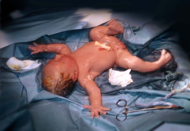
Image: “Unidentified newborn infant moments after the umbilical cord had been cut. Photo taken by Every1blowz, August 25, 1994.” by Mad Max at the English language Wikipedia. License: CC BY-SA 3.0
Definition of the Third Stage of Labor
The third stage of labor is generally the fastest stage of labor, lasting less than five minutes in an uncomplicated delivery. The infant has been delivered and there is still some minor bleeding.
Inspection for perineal tears and their repair
Episiotomies and lacerations are evaluated and repaired before the separation of the placenta if possible to allow for visualization before blood gushes out due to placental separation.
There are several types of episiotomies. Most episiotomies are midline, meaning the cut is made straight down from the vaginal introitus toward the rectum. In cases where more room is needed, a mediolateral episiotomy can be done but this is more difficult to repair and causes more post-procedure pain.
In a midline episiotomy, the perineal tear/lacerations are classified as:
These are repaired by repairing the anus and anal sphincter first, followed by repair of the perineal muscles. Absorbable sutures 2-0 or 3-0 are used. When these are approximated, the vulvar tissues and vaginal epithelium are approximated, and the episiotomy can heal with little risk of infection or secondary complications.
Delivery of the placenta
The uterus contracts to expel the placenta, which ideally is intact with no remaining placental tissue in the uterus. This should take two minutes but may take as long as 30 minutes.
Signs of placental separation include:
- There is a gush of fresh blood from the vagina.
- Umbilical cord lengthens.
- Fundus of the uterus rises.
- Uterus becomes firm and globular.
Symptoms and Signs of the Third Stage of Labor
The third stage of labor is marked by a significant decline in pain. The mother is generally distracted by the delivery of her infant and pays little attention to the third stage of labor. The woman will continue to have cramping in the lower abdomen and pelvis as the uterus contracts to expel the placenta. If the woman has had epidural anesthesia, this is continued so any lacerations or episiotomies can be repaired.
The third stage of labor presents with an umbilical cord remaining in the vaginal opening. The perineum may be intact or may have suffered some lacerations. Routine episiotomy is no longer recommended for normal vaginal deliveries but may be done when expeditious delivery is necessary in the third stage of labor. Lacerations or episiotomies can begin to be repaired before the placenta presents itself.
Signs that the placenta has detached include elongation of the umbilical cord, a change in shape of the uterus so that it is more globular, and a sudden gush of blood that signals the detachment of the placenta from the uterine wall.
The doctor or midwife can facilitate the expulsion of the placenta by putting downward pressure in the suprapubic area and gently tugging on the cord. Failing to put downward pressure in the suprapubic area may cause inversion of the uterus if too much traction is applied to the placenta. In some cases, manual extraction of the placenta may happen before the placenta disengages.
Special Tests in the Third Stage of Labor
No special tests need to be done in a normal delivery. In cases of severe postpartum hemorrhage of unknown etiology, tests of clotting function, such as a bleeding time, prothrombin time, and partial thromboplastin time may be ordered to identify an underlying bleeding pathology in the mother.
Complications in the Third Stage of Labor
The only complications of the third stage of labor are ;
- Retained placenta or retained placental fragments
- Tears into the perineum or rectum that need to be repaired to restore the anus and perineal tissues, so they are approximated with dissolvable sutures.
- Postpartum hemorrhage
If placental delivery and perineal tear repair are done correctly, hemorrhage rarely persists. Moreover, a labor and delivery nurse should continuemassaging of the uterus to maximize the tone of the uterus. And encourage contraction and arrest of bleeding. Postpartum hemorrhage is seen in cases of retained placental fragments, atony of the uterus, laceration, and a preexisting bleeding disorder.
Primary postpartum hemorrhage occurs in the first 24 hours after delivery. Secondary postpartum hemorrhage occurs 24 hours to 12 weeks after delivery. Postpartum hemorrhage is defined by blood loss:
- > 500 cc for vaginal delivery
- > 1,000 cc for a cesarean delivery
Common indicators of a likely post-partum hemorrhage event include:
- Prolonged labor/previous C-S/ polyhydramnios
- Antepartum hemorrhage.
- Recent history of bleeding.
- Uterine fibroids.
- Multiparity.
Treatment in the Third Stage of Labor
There are two main interventions that need to be done in the third stage of labor. The first is the facilitation of the evacuation of the placenta described above. If this is done correctly, continued massage of the uterus is done by a labor and delivery nurse to maximize the tone of the uterus.
If there is ongoing uterine atony, the patient may be given any one of four drugs to increase uterine tone. Additional intravenous oxytocin may be given to increase tone. Methylergonovinecan be given if there is no maternal hypertension. PGF2a can be given if there is no maternal asthma. Misoprostol can be given without any contraindications.
If uterine massage and medications fail to stop the bleeding, surgical interventions may be considered. The patient may be taken to the operating room to have a curettage to remove placental fragments. The cervix and perineum need to be carefully examined for an extrauterine cause of the bleeding.
If the curettage fails and there is ongoing hemorrhage not caused by a bleeding disorder, a uterine artery embolization may be necessary. Packing of the uterus may stop the bleeding. Arterial ligation may control hemorrhage. In severe cases, where nothing is stopping the bleeding, the patient may need an emergency hysterectomy.
If there is evidence for a bleeding disorder as the cause of the bleeding, the patient may need blood products to replace lost blood. Packed red blood cells can be given along with fresh frozen plasma, platelets, and cryoprecipitate as the cause of the bleeding disorder may be unable to be ascertained in the short period of time necessary to control the bleeding.
Episiotomy repair is the second intervention that needs to happen. There are several types of episiotomies. Most episiotomies are midline, meaning the cut is made straight down from the vaginal introitus toward the rectum. In cases where more room is needed, a mediolateralepisiotomy can be done but this is more difficult to repair and causes more postprocedure pain.
In a midline episiotomy, a first-degree laceration can happen, which involves a tear into the vulva and vaginal epithelium. A second-degree laceration involves a tear into the perineal muscles with an intact anal sphincter. A third-degree laceration involves a tear involving the anal sphincter. A fourth-degree tear involves a tear into the mucosa of the anus.
These are repaired by repairing the anus and anal sphincter first, followed by repair of the perineal muscles. When these are approximated, the vulvar tissues and vaginal epithelium are approximated and the episiotomy is allowed to heal with little risk of infection or secondary complications.
Prognosis of the Third Stage of Labor
The prognosis is excellent in the third stage of labor. The risk of postpartum hemorrhage when there is active management to control bleeding is about 5 %. Care must be taken to remove the placenta without causing inversion of the uterus, and the anus, perineum, and cervix must be examined for lacerations or hematomas.
If lacerations are present, these are sutured with absorbable sutures to control bleeding and restore function to the anal and perineal structures.
Review Questions
The correct answers can be found below the references.
1. You are treating a multiparous woman who has had two normal vaginal deliveries in the past. The third stage of labor is complicated by hemorrhaging after removal of the placenta. What do you suspect is the cause of the bleeding?
- Bleeding diathesis in the mother
- Retained placental fragments
- Uterine atony
- Inverted uterus
2. You are caring for a primiparous woman who required an episiotomy and who sustained a fourth-degree laceration of her midline episiotomy. How do you go about repairing the tear?
- Repair the anal sphincter first and then repair the perineal muscles.
- Repair the anal epithelium first and then repair the anal sphincter before approximating the perineal tissues.
- Approximate the vaginal mucosa only as the muscles will approximate themselves.
- Repair the perineal muscles and then the anal sphincter. Follow this by repairing both the anal epithelium and the vaginal epithelium.
3. You are caring for a woman who has retained placental fragments. She is bleeding moderately but is hemodynamically stable. What is your next step?
- Manually remove the placental fragments at the bedside.
- Perform a uterine artery ligation.
- Do a dilatation and curettage in the operating room.
- Give misoprostol to increase uterine tone so the fragments can be expelled.

Comentários
Enviar um comentário