Stomach Ulcer (Peptic Ulcer) — Symptoms and Treatment
Table of Contents
- Definition and Background of Peptic Ulcer
- Pathophysiology of Peptic Ulcer
- Etiology of Peptic Ulcer
- Epidemiology of Peptic Ulcer
- Presentation of a Peptic Ulcer
- Classification of a Peptic Ulcer
- Differential Diagnoses of Peptic Ulcer Disease
- Diagnosis of Peptic Ulcer Disease
- The Management of Peptic Ulcers
- Review Questions
- References
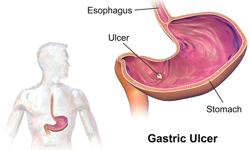
Image: “Gastric Ulcer” by BruceBlaus. License: CC BY-SA 4.0
Definition and Background of Peptic Ulcer
Open sores are called ulcers, and the word peptic means that acid is the cause of the ulcer. Gastric and duodenal ulcers are the two most common types of peptic ulcers. The difference between the two types is in the location of the ulcer. Duodenal ulcers are found in the duodenum, and gastric ulcers are located in the stomach.
Pathophysiology of Peptic Ulcer
There is a physiologic balance in normal conditions between gastroduodenal mucosal defenseand gastric acid secretion. When this balance between the defensive mechanisms and the aggressive factors gets disrupted, mucosal injury occurs, which results in the developing of peptic ulcer. These aggressive factors include H. pylori infection, NSAIDs, alcohol, smoking, acid, bile salts and pepsin.
Etiology of Peptic Ulcer
H. pylori infection
H. pylori infection is considered as one of the most common causes of peptic ulcer disease. The prevalence of H. pylori in complicated ulcers is less than that in uncomplicated ulcers.
Drugs
The use of nonsteroidal anti-inflammatory drugs (NSAID) is a common cause for the developing of peptic ulcer disease. They render the mucosa vulnerable to injury by disrupting the mucosal permeability barrier. Other risk factors, when accompanied with NSAID use, increase the risk of developing peptic ulcers; these factors include advanced age, female sex, history of a previous peptic ulcer, long-term use or high doses of NSAID and severe comorbid diseases.
Lifestyle factors
Smoking tobacco and drinking alcohol appears to increase the risk of developing peptic ulcer disease. There is not enough evidence that the use of caffeine increases the risk of developing ulcers.
Severe physiologic stress
Several stressful conditions may cause peptic ulcers such as CNS trauma, surgery, burns, severe medical illness, sepsis, hypotension, multiple traumatic injuries and respiratory failure.
Hypersecretory states
Several hypersecretory states may cause peptic ulcers including:
- Systemic mastocytosis
- Basophilic leukemias
- Short bowel syndrome
- Gastrinoma
- Hyperparathyroidism
- Cystic fibrosis
Genetic factors
Approximately 20% of peptic ulcer disease patients have a family history of the disease. Studies have also shown a weak association between blood type O and the development of duodenal ulcers.
Epidemiology of Peptic Ulcer
Approximately 4.5 million people are affected with peptic ulcer disease every year in the United States with nearly 10% of the US population showing evidence of a duodenal ulcer at some time in their lives.
The frequency of peptic ulcer disease in other countries is variable and is determined by the association with the major risk factors of the condition, such as NSAID use and H. pylori infections.
Presentation of a Peptic Ulcer
Obtaining a thorough medical history is essential in the diagnosis of peptic ulcer disease; however, history alone is not enough to reach a definite diagnosis.
Symptoms of peptic ulcer
The most common symptom of both duodenal and gastric ulcers is an epigastric pain, which is characterized by a burning sensation occurring after meals. In gastric ulcers, the pain usually starts shortly after meals; whereas, it may take a few hours to start in duodenal ulcers. Unlike duodenal ulcers, the pain caused by gastric ulcers cannot be relieved by antacids.
Other symptoms and manifestations include:
- Heartburn
- Chest discomfort
- Dyspepsia
- Hematemesis
- Fatigue
- Dyspnea
- Hematochezia may present in rare cases of bleeding ulcers
- Peptic ulcer disease may be asymptomatic, especially in elderly patients
Alarm features that require referral to a gastroenterologist and further workup include:
- Unexplained weight loss
- Recurrent vomiting
- Family history of GI cancer
- Progressive dysphagia or odynophagia
- Anemia or bleeding
- Early satiety
Physical examination signs of a peptic ulcer
The physical findings in uncomplicated peptic ulcer disease are nonspecific, and they include:
- Mild epigastric tenderness
- Melena, which results from acute or subacute GI bleeding
- Succussion splash resulting from complete or partial gastric obstruction
- Occult blood loss results in guaiac-positive stool
If the peptic ulcer perforates, patients will suffer from a sudden, severe sharp abdominal pain. Generalized and rebound tenderness, as well as guarding and rigidity, will be revealed by physical examination.
Classification of a Peptic Ulcer
The Johnson classification categorizes gastric ulcers into four types:
Type I
Type I gastric ulcers are located on the lesser curvature near the angularis incisura, which is close to the border between the body of the stomach and the antrum. Acid secretion is usually normal or decreased in patients suffering from type I gastric ulcers.
Type II
They are a combination of duodenal and stomach ulcers. Acid secretion is increased in these types of ulcers.
Type III
These ulcers are prepyloric. Acid secretion in type III ulcers could be normal or increased.
Type IV
Ulcers in this type are usually located near the gastroesophageal junction. The acid secreted is either normal or decreased in this type.
Differential Diagnoses of Peptic Ulcer Disease
- Crohn’s disease
- Zollinger-Ellison syndrome
- Acute gastritis
- Acute coronary syndrome
- Cholecystitis
- Acute cholangitis
- Diverticulitis
- Chronic gastritis
- Esophagitis
- Esophageal rupture
- Gallstones
- Gastroesophageal reflux diseases
- Viral hepatitis
- Inflammatory bowel disease
Diagnosis of Peptic Ulcer Disease
H. pylori testing
H. pylori infection is one of the most common causes of peptic ulcer disease. Thus, it is important to test for it. Usually, routine laboratory tests are not helpful; endoscopic and radiographic confirmation is required for the documentation of peptic ulcer disease.
Endoscopy
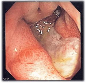
The preferred diagnostic test in the evaluation of suspected peptic ulcer patients is upper GI endoscopy. It is highly sensitive, helps in the detection of H. pylori infection, and aids in the differentiation between benign and malignant lesions through biopsies.
Image: “Endoscopic image of deep gastric ulcer in the gastric antrum” by Samir. License: CC BY-SA 3.0
Radiography
Chest radiographs are used to look for abdominal air to detect ulcer perforation.
Angiography
Patients with massive GI bleeding, who cannot undergo endoscopy, may benefit from angiography, which helps in identifying the source of bleeding and providing the needed therapy.
Serum gastrin level
Screening for Zollinger-Ellison syndrome is performed by a fasting serum gastrin level.
Secretin stimulation test
If the serum gastrin level test was not enough to make the diagnosis of Zollinger-Ellison syndrome, a secretin stimulation test may be used, which distinguishes Zollinger-Ellison syndrome from other conditions.
Biopsy
A biopsy is taken to examine for gastric cancer, which has a 70% accuracy. The sensitivity increases to 99% when seven biopsy samples are obtained from the base and ulcer margins.
Histologic findings
Slough and inflammatory debris cover the surface of the ulcer. Active granulation with fibrinoid necrosis and mononuclear leukocytic infiltration may be seen beneath the neutrophilic infiltration. The mucosa and submucosa are usually infiltrated by plasma cells, monocytes, and lymphocytes in chronic superficial gastritis.
The Management of Peptic Ulcers
The management of peptic ulcers differs depending on the clinical presentation, as well as the etiology of the condition.
Emergency department cares in peptic ulcer
In the emergency department, the presentations of gastritis and peptic ulcer disease are indistinguishable, which makes the management of them the same. Relieving the discomfortand promoting healing by the protection of the gastric mucosal barrier are the main treatment goals in the management of acute conditions. Acute interventions are usually not needed in peptic ulcer disease or gastritis.
H. pylori infection
H. pylori infections are treated with triple therapy including amoxicillin, clarithromycin and a PPI for 7 to 14 days. In patients who are allergic to penicillin, amoxicillin is replaced with metronidazole. It is recommended to extend the duration of the treatment for more than 14 days in complicated cases of H. pylori infections.
Medical management of NSAID ulcers
It is advised to discontinue the use of NSAID in patients who are diagnosed with peptic ulcer disease.
Bleeding peptic ulcers
A bleeding ulcer can be evaluated and managed by endoscopy, which reduces the likelihood of recurrent bleeding and decreases the need for surgical intervention.
The general pharmacologic principle of the medical management of acute bleeding from a peptic ulcer is acid suppression.
Review Questions
The answers are below the references.
1. A 51-year-old Caucasian man presented to the doctor’s office complaining of a black tarry stool. The development of carcinoma in this patient is least associated with which of the following causes?
- Adenomatous polyp
- Duodenal ulcer
- Barrett’s esophagus
- H. pylori infections
- Gastric ulcer
2. A 32-year-old Nigerian woman presents to your office complaining of a burning pain in her epigastric region. An ulcer in the proximal duodenum was demonstrated by esophagoduodenoscopy. The cause of this patient’s ulcer is best addressed by which of the following treatments?
- Surgical resection
- Omeprazole
- Antibiotics
- NSAID use cessation
- Cimetidine
3. A 33-year-old Caucasian man presented to your office complaining of symptoms suggestive of peptic ulcer disease. You decide to perform a biopsy because you suspect H. pylori infection. Which site of the stomach (based on the figure on the right) corresponds to the likely location of the bacterium?
- Lower esophageal sphincter
- Pyloric sphincter
- Antrum of the stomach
- Cardia
- Fundus


It's a nice article. Everyone should read. Thanks for sharing. For more amazing information you may get from this link
ResponderEliminarpeptic ulcer
Am Richard, I am here to testify about a great herbalist man who cured my wife of breast cancer. His name is Dr Imoloa. My wife went through this pain for 3 years, i almost spent all i had, until i saw some testimonies online on how Dr. Imoloa cure them from their diseases, immediately i contacted him through. then he told me the necessary things to do before he will send the herbal medicine. Wish he did through DHL courier service, And he instructed us on how to apply or drink the medicine for good two weeks. and to greatest surprise before the upper third week my wife was relief from all the pains, Believe me, that was how my wife was cured from breast cancer by this great man. He also have powerful herbal medicine to cure diseases like: Alzheimer's disease, parkinson's disease, vaginal cancer, epilepsy Anxiety Disorders, Autoimmune Disease, Back Pain, Back Sprain, Bipolar Disorder, Brain Tumor, Malignant, Bruxism, Bulimia, Cervical Disc Disease, Cardiovascular Disease, Neoplasms , chronic respiratory disease, mental and behavioral disorder, Cystic Fibrosis, Hypertension, Diabetes, Asthma, Autoimmune inflammatory media arthritis ed. chronic kidney disease, inflammatory joint disease, impotence, alcohol spectrum feta, dysthymic disorder, eczema, tuberculosis, chronic fatigue syndrome, constipation, inflammatory bowel disease, lupus disease, mouth ulcer, mouth cancer, body pain, fever, hepatitis ABC, syphilis, diarrhea, HIV / AIDS, Huntington's disease, back acne, chronic kidney failure, addison's disease, chronic pain, Crohn's pain, cystic fibrosis, fibromyalgia, inflammatory Bowel disease, fungal nail disease, Lyme disease, Celia disease, Lymphoma, Major depression, Malignant melanoma, Mania, Melorheostosis, Meniere's disease, Mucopolysaccharidosis, Multiple sclerosis, Muscular dystrophy, Rheumatoid arthritis. You can reach him Email Via drimolaherbalmademedicine@gmail.com / whatsapp +2347081986098
ResponderEliminar