Mitral Stenosis (Mitral Valve Stenosis) — Symptoms and Treatment
Table of Contents
- Introduction
- Definition of Mitral Stenosis
- Etiology of Mitral Stenosis
- Classification of Mitral Stenosis
- Pathophysiology of Mitral Stenosis
- Symptoms and Clinical Presentation of Mitral Stenosis
- Progression and Special Types of Mitral Stenosis
- Diagnosis of Mitral Stenosis
- Treatment of Mitral Stenosis
- Complications of Mitral Stenosis
- References
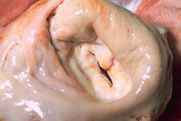
Image: „Gross pathology of heart showing mitral stenosis” by CDC. Licence: Public Domain
Introduction
Mitral valve stenosis is, in most cases, a long-term sequel of rheumatic fever. The severity may depend on the remaining open area and determines which symptoms occur.
Definition of Mitral Stenosis
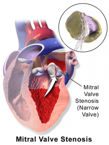
Mitral valve stenosis refers to a narrowing of the mitral valve, which obstructs the filling of the left ventricle.
Etiology of Mitral Stenosis
Causes of Mitral Stenosis
The most common cause of mitral valve stenosis is rheumatic fever, which, however, may date back many years. Therefore, asking the patient about the history of frequent bacterial tonsillitis is important. Rarely, there may be congenital stenosis in the mitral valve area.
Classification of Mitral Stenosis
The 3 Grades of Severity of Mitral Stenosis
Depending on the remaining open area, mean pressure gradient and mean pulmonary capillary pressure, mitral valve stenosis can be divided into the severity grades of mild, moderate and severe. The mild grade has a pressure gradient of less than 7 mmHg, an opening area that is between 1.5 and 2 cm2, and a mean pulmonary capillary pressure of less than 20 mmHg.
The moderate grade has a mean pressure gradient of 8 – 15 mmHg, an opening area of 1 to 1.5 cm2, and a mean pulmonary capillary pressure of 21 – 25 mmHg. In severemitral stenosis, the mean pressure gradient increases to more than 15 mmHg, the opening area drops below 1 cm2and the mean pulmonary capillary pressure rises to more than 25 mmHg.
Pathophysiology of Mitral Stenosis
What Happens in Mitral Stenosis?
Because of the stenosis of a mitral valve, the blood cannot flow properly into the left ventricle. Thus, this causes a blood stasis in the left atrium, and the blood flows back into the lungs to the pulmonary veins, and ultimately in the right heart. Therefore, there is a risk of right heart failure and an increased risk of atrial fibrillation due to the overstretching of the left atrium. Atrial fibrillation may increase the risk of embolization and may result in acute ischemia of the legs, stroke or mesenteric ischemia
Symptoms and Clinical Presentation of Mitral Stenosis
The severity of the disease determines which symptoms occur. The expansion of the left atrium may lead to atrial fibrillation and thrombi, and therefore to arterial embolisms. Due to the congestion of the blood in the lungs, this leads to pulmonary hypertension with symptoms such as dyspnea and nocturnal cough. Signs of right heart failure may also occur.
The reduced cardiac output leads to:
- Performance degradation
- Fatigue
- Peripheral cyanoses
A late finding and a bad prognostic factor mean that the right ventricle can no longer keep up with the increased pressure transmitted backward from the LA.
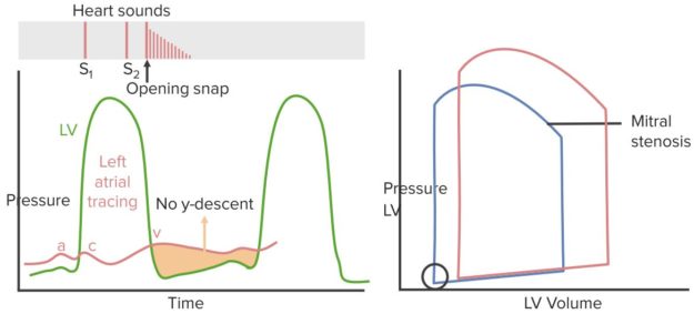
Physical examination will reveal palpation. Right ventricular heave if patient has pulmonary hypertension; PMI is normal or decreased. Physical examination will also reveal mid-diastolic murmur. Remember: blood flows across the mitral valve and into the LV during diastole.
Heard loudest at the mitral area (apex) and has a “rumbling” characteristic. Radiates toward the axilla and an opening “snap” as the calcigic valve is forced open by the LA contraction. Atrial fibrillation may be a consequence of the dilated LA.
Progression and Special Types of Mitral Stenosis
The progression of mitral valve stenosis depends on the severity. Pulmonary edema and right heart failure are the leading causes of death. Arterial and pulmonary embolisms are also complications that are potentially fatal.
Diagnosis of Mitral Stenosis
Auscultation of Mitral Stenosis
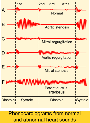
During auscultation, a loud first heart sound, as well as a split second heart sound and a mitral opening snap, can be heard. A mid-diastolic rumbling murmur with can be best heard over the cardiac apex. A sign of reactive pulmonary regurgitation is the Graham Steel murmur. A presystolic crescendo murmur can be heard.
“Mitral stenosis” Image created by Lecturio
Radiological Examination of Mitral Stenosis
An ECG may show a P with double peaks, as well as a right ventricular hypertrophy mark, and a right axis deviation. A radiograph may also show a right heart enlargement, as well as an enlarged left atrium, and a mitral configuration. During echocardiography, the valve can be readily assessed and a quantification of the grade of stenosis can be made. In addition, a cardiac catheterization can be performed for evaluation.
Treatment of Mitral Stenosis
Treatment Options for Mitral Stenosis
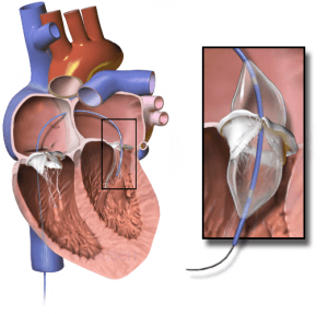
Conservative treatment options include drug treatment of heart failure and maintaining a normal frequency sinus rhythm, as well as thromboembolism and endocarditis prophylaxis. The catheter method offers the possibility of a mitral valve valvuloplasty, which can help avoid a large surgical intervention.
Surgical repair or replacement of the mitral valve if the patient stays symptomic despite medical treatment. Mitral valve replacement is the surgical treatment approach, which is indicated with a moderate stenosis or with a failure of valvuloplasty.
Atrial fibrillation occurring as a complication of mitral stenosis should be treated. Anticoagulation treatment and consider restoration of NSR. Treatment of fluid overload with diuretics should be done.
Complications of Mitral Stenosis
Complications of mitral stenosis include arterial embolism, bacterial endocarditis and pulmonary edema.
Comentários
Enviar um comentário