Nursemaid´s Elbow (Radial Head Subluxation) — Signs and Radiology
Table of Contents
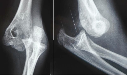
Image: “Radiographs showing a posterolateral dislocation of the elbow. (A) Anteroposterior view. (B) Lateral view.” by Martín JR, Mazzini JP. License: CC BY 3.0
Definition
Nurse maid’s elbow is also known as pulled elbow due to its etiologic mechanism. The medical term for the mechanism is Radial head subluxation.
Epidemiology
It is a common problem in the pediatric population representing the most common form of upper limb injuries for children under the age of 6 years. It has been recorded in patients aged 4 – 31 years. The peak number of cases of Nursemaid’s elbow occurs at 26 months of age.
It has a slight female predominance with the left hand being affected in most times.
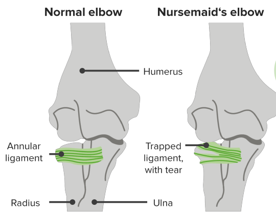
Anatomy of Nursemaid´s Elbow
The elbow is made up of two joints:
- The humeroulnar joint that joins the humerus with the ulna and allows the bending of the elbow.
- The radiocapitellar joint that holds the radius and humerus, so the forearm can be rotated into the supine and prone position.
Discussion of Nursemaid´s Elbow
It is a common problem in the pediatric population. The elbow gets slipped out of the normal joint space. Radius (elbow bone) is attached to the humerus through elastic ligaments. These ligaments gain strength and flexibility with the passage of time.
In children, the ligaments are weak so a detachment can easily occur, leading to Nursemaid’s elbow. It can happen with a mild force such as swinging the babies while holding their arms. The condition is also known as ‘pulled elbow’ or ‘radial head subluxation.’
Toddlers and pre-schoolers mostly get the injury as the tissues are softer and liable to lacerations. After 5 or 6 years of age, the incidence gets reduced as the ligaments become stiffer and tighter. Girls are more prone to this condition. The children aged 1–4 years are more susceptible, and infants from 6–12 months of age. The peak number of cases of Nursemaid’s elbow occurs at 26 months of age.
Anatomy of Nursemaid´s Elbow
The elbow is made up of two joints – one is a humeroulnar joint and the other is radiocapitellar joint. The humeroulnar joint joins the humerus with the ulna and allows the bending of the elbow. The radiocapitellar joint holds the radius and humerus so the forearm can be rotated into the supine and prone position.
In Nursemaid’s elbow, it is the radiocapitellar joint which gets partially separated. The annular ligament which surrounds the radial bone is responsible for Nursemaid’s elbow. The ligament is weaker in the young that can be easily disrupted with a minor force.
Causes of Nursemaid´s Elbow
The condition can occur as a result of the following reasons:
- Lifting the child by the hands or wrists
- Stopping the fall of a baby by grabbing the arms
- Swinging the child by the arms
- The infant rolling over the arm
- During the fall on an outstretched hand from a height
- In the case of child abuse
Signs and Symptoms of Nursemaid’s elbow
It is an acute condition that occurs after the injury. The child tends to hold the arm by the side, the elbow is slightly bent and ventral surface of hand is facing towards the body. The left arm is said to be more commonly injured. The various signs and symptoms include:
- Pain and tenderness on a lateral aspect of the elbow.
- Swelling and bruising can also be the rare presentation.
Diagnosis of Nursemaid´s Elbow
The diagnosis is clinical, based on history and examination. Usually, the radiological investigations like X-rays, US, CT scans or bone scans are not required. These can be carried out to rule out other suspected fractures.
Management of Nursemaid´s Elbow
The treatment is based on the age and medical history of the child. It is a temporary condition but can yield permanent consequences if not timely treated.
Emergency treatment may be needed to immobilize the fracture to avoid further harm and control pain. This protection can also be carried out in the prehospital care setup.
Medical Treatment
Analgesics and anti-inflammatory medicines are prescribed to alleviate the pain.
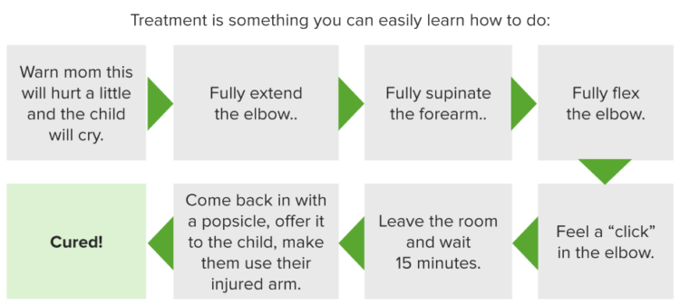
Non-operative maneuvers
This involves closed reduction, which is indicated in the acute cases. This procedure can be performed on an outpatient basis. It is painful and a child may cry for few minutes. The closed reduction can be done within a few minutes by following these steps:
- The forearm is supinated and the elbow is flexed to more than 90 degrees.
- The thumb is placed over the radial head and a click sound is produced when it gets reduced.
- Wait for few minutes.
- Allow the child to use the injured arm.
- Follow-up is required if the child cannot resume the normal movements.
Sometimes, the first attempt does not succeed, so two or three may be needed to correct it.
Operative
This includes the open reduction and fixation under proper anesthesia. Mostly used for the chronic cases of Nursemaid’s elbow.
Prognosis
The condition runs a predictable course as the deformities are not permanent and no injury results from the condition. However, recurrence may be seen in approximately 25% of those who suffer from the condition at a younger age of <2 years.


Comentários
Enviar um comentário