Maxillary Bone and Le Fort Fractures
Table of Contents
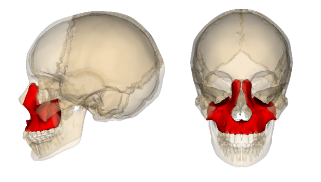
Image: “Maxilla”, generated by Life Science Databases (LSDB). License: CC BY-SA 2.1 JP
The Facial Bones
The human skull is made up of the following bones in the face :

Image: “Anterior view of the facial bones” by Polygon data is from BodyParts3D License: CC BY-SA 2.1 JP
- Inferior nasal concha (2) – bright green
- Lacrimal bones (2) – bright blue
- Mandible (1) – purple
- Maxilla (2) – yellow
- Nasal bones (2) – pink
- Palatine bones (2) – red
- Vomer (1) – blue
- Zygomatic bones (2) – dark green
Facial Fractures
The maxilla, mandible, and zygomatic bones are the major bones of the face. Fracture of the facial bones can lead to permanent deformitiesand can be very life threatening. Most patterns of facial fractures involve the maxilla. Facial fracture patterns include:
- Tripod fracture
- Le Fort I (horizontal or floating palate)
- Le Fort II (pyramidal)
- Le Fort III (transverse)
- fractures of the cheekbone.
- Alveolar processes of the maxilla
- Smash fractures
Nasal bones: this is often known as broken nose. The nose is made up of tiny bones which require little force to break compared to other facial bones. This leads to deformation as well as swelling of the nose.
Orbital fractures: this entails fractures to the eye sockets and are classified into 3 type:
- Orbital rim fracture: this is fracture to the outer rim of the eye socket. Despite being the thickest, damages might result into optic nerve breakdown.
- Direct Orbital floor fracture: this type of fracture extends to the lower part of the eye socket
Blowout fractures: this type of fracture results into a crack of the lower part of the eye sockets however the orbital rim remains intact.
Cause of Facial Fractures
Like most fractures, the cause is blunt force trauma. This commonly occurs during road traffic accidents (RTA), steering wheel injuries, falls, and violent attacks/fighting. During RTA, facial trauma is usually not the only injury that occurs, it is commonly accompanied by chest trauma and other head injury.
Le Fort Fractures
René Le Fort studied the various types of fractures involving the facial bones and created a classification system for certain types of these fractures – they are now referred to as Le Fort fractures.
Le Fort fractures involve separation of all or a portion of the midface from the skull base. Force on the face result into fractures along lines of weaknesses in the mid-face. One of the key components of these types of fractures is the involvement of the pterygoid plate. The system is based on the level of injury with respect to the maxilla – the higher the Le Fort classification, the more superior the fracture. The common characteristic with all types of the Le Fort fractures is that they are pterygoid processes along the sphenoid bone. There are 3 classes of Le Fort fracture:
Le Fort type 1

Image: “Le Fort fractures – types” by RosarioVanTulpe, License: CC BY-SA 3.0
This type is also called the “floating palate.” Usually the result of a downward blow to the upper jaw. The fracture line passes superior to the maxillary alveolar processes, separating the teeth from the upper face.
It involves the alveolar ridges, lateral part of the nose, body of the nasal septum and inferior part of the maxillary sinus. Lower maxilla and the maxillary teeth are the floating fragments.
Le Fort type 2
The fracture of the face has a pyramidal shape. Usually results from a blow to the middle to lower maxilla. The key component is involvement of the inferior orbital rim. At the base of the pyramid lie the teeth, apex of the pyramid is formed by the nasofrontal suture.
Fracture line passes from the posterior alveolar ridge then the lateral walls of maxillary sinuses and inferior rim of the orbit till the nasal bones. This type of fracture involves the posterolateral wall of the maxillary sinus and the infraorbital foramina. Lacrimal or ethmoid bones can also be fractured along with the maxilla.
Le Fort type 3
These are transverse fractures, also known as craniofacial dissociation. Can result from a blow to the nasal bridge or upper maxilla. These fractures involve the zygomatic arch, the orbital wall and floor, ethmoids, vomer, and nasofrontal suture. The fracture line is in the horizontal plane and passes through the superior orbital fissure. The facial bones can be massively disturbed. Figure shows the fracture line passing through the above mentioned structures. A frequent symptom of these fractures is cerebrospinal fluid (CSF) rhinorrhea (leakage through the nose).
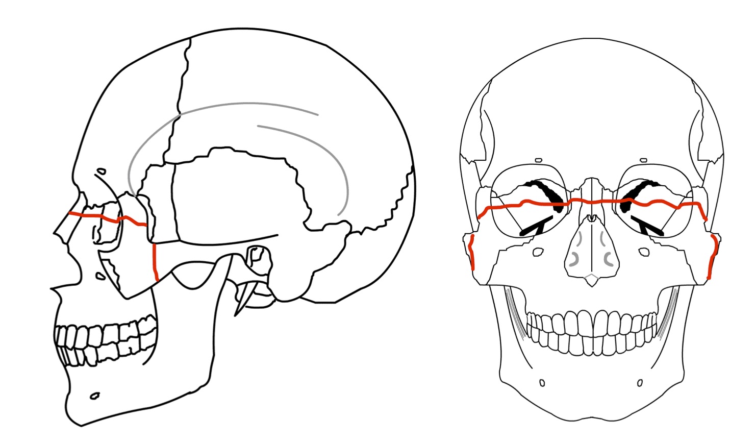
Image: “Le Fort type 3 lateral and anterior view” by RosarioVanTulpe, License: CC BY-SA 3.0
Associated Injuries with Le Fort Fractures
Le Fort fractures of the midface can occur in combination as well. Any combination of these three types of fractures is possible. Occasionally, one type of fracture occurs on one side and another type occurs on the other. Complications associated with these type of fractures include airway disturbance, loss of consciousness, intracranial injury, vision and/or hearing changes, and cranial nerve injuries.
Important points in the classification of mid facial fractures are given below:
- If the lateral or anterolateral margin of the nasal fossa remains intact after trauma or injury, it is not classified as a Le Fort type 1.
- If the fracture line passes through the posterior alveolar ridges and lateral wall of the maxillary sinus, but infraorbital margin remains intact, it is not classified as a Le Fort type 2.
- It is not a Le Fort type 3 if the zygomatic arch remains intact, even if the fracture involves the other structures.
Evaluation of Patient with Maxillary Fractures
After a trauma, if the patient has been removed from any life threatening situation and possible airway and/or bleeding problems have been addressed properly, a detailed examinationshould be conducted to help rule out any facial/skull fracture. Soft tissues produce various changes like swelling, ecchymosis, blood clots, and gross blood loss. Hematomas usually occur at the site of fracture. Periorbital swelling more commonly occurs in type 2 and type 3 Le Fort fractures. Open bite deformity can also occur with these fractures.
Changes in vision can represent injury to the optic canal, the eye, or even the retina. Depending on the pattern of injury and symptoms present, it may be warranted to request a consult from ophthalmology, oromaxillofacial surgery, anesthesiology/respiratory, eye-ear-nose-throat, and/or radiology.
Note: The maxillary fracture accounts for about 6-25% of all facial fractures.
Overall examination
- Examination of the cranium and skull – are there signs of trauma/bleeding?
- Examination of the face – swelling/deformity?
- Examination of the eyes and orbit – swelling/hemorrhage/ecchymosis?
- Examination of the oral and nasal cavity with the airway – is there clear rhinorrhea/epistaxis?
- Examination of the mouth/jaw/teeth – deformity/inability to close mouth/pain with tapping of upper teeth?
- Evaluation of hearing/ear examination – is there battle sign or blood/CSF leakage through ears?
Investigation used in Le Fort fractures

Image: “X-ray lateral view showing nasal bone fracture” by © Nevit Dilmen, License: CC BY-SA 3.0
X-ray of the face from different views is taken to check the bone symmetry and fractured bony parts. Waters view, Caldwell view, lateral view and occasionally submentovertex view are the various X-rays taken.
Nasal bone fracture is viewed on lateral X-ray. Cervical films should also be taken to ensure the normal structures of the cervical spine.
High-resolution CT scan of the head and face is the investigation of choice for the facial fractures. MRI can also be done to assess the soft tissue injuries.
Radiological signs for the facial fractures
- Non-anatomic linear lucency
- Cortical defect or disturbance of the suture line
- Bony fragments overlapping “double-density” seen on the imaging study
- Asymmetry of facial bones
- Soft tissue swelling
- Periorbital or intracranial air with signs of edema
- Fluid in a paranasal sinus or hematoma in the epidural space
Management of Facial Fractures
These fractures come in the domain of orthopedic and oromaxillofacial surgery. Relevant surgery according to the nature of the injury is done after maintaining airway and circulation. If a dislodged bone obstructing the airway is suspected, tracheostomy should be considered before any surgical procedure is done. Severe bleeding can occur from intranasal structures or lacerations due to dislodged bony fragments. Bleeding can be stopped by pressure packing, cauterization, and/or suturing of the wound.
For unstable floating fragments, surgical fixation is indicated to restore normal function and anatomical symmetry.
How facial fractures can be prevented
It is worth noting that there are no methods that can aid in completely preventing the fracture to the face, however, the following measures can be put in place in order to minimize the extent of damage associated with the facial fractures:
- Wearing protective gears such as helmets as well as seat belts while driving
- While playing sport ensure that you have face masks and helmets
- Strictly follow the safety guidelines within your working environment


Comentários
Enviar um comentário