Pityriasis Rosea (Pityriasis Rosea Gibert) in Children — Symptoms and Treatment
Table of Contents
- Definition
- Epidemiology of Pityriasis Rosea in Children
- Pathophysiology of Pityriasis Rosea
- Clinical Presentation of Pityriasis Rosea in Children
- Diagnostic Workup for Pityriasis Rosea in Children
- Differential Diagnosis
- Treatment of Pityriasis Rosea in Children
- Prognosis of Pityriasis Rosea
- References
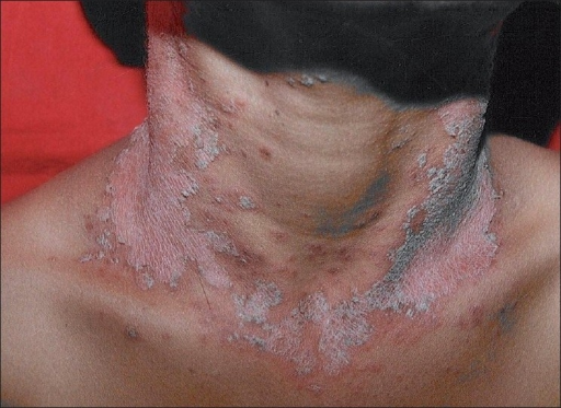
Image: “Sudden appearance of scaly sharply demarcated erythematous lesions on front of neck in the next week” by Zawar V. License: CC BY0 2.0
Definition
Pityriasis rosea (PR) is a fine pinkish scaly rash that is benign. The rash is characterized by being acute, self-limiting, and papulosquamous. The skin lesions usually begin with a herald patch and evolve quickly. Whether the condition has a viral etiology is still debatable.
Epidemiology of Pityriasis Rosea in Children
PR is a quite common condition in children with an estimated annual incidence of 0.13%. Seasonal variety in the incidence of PR has been noted which agrees with the possibility of a viral etiology of the condition. The condition is more common in the spring and winter seasons.
Another point in favor of an infectious etiology for PR is the observation that a higher incidence, 2%, has been documented in the developing world. This, however, can be also attributed to the hot weather that is observed in India and Malaysia, rather than a true infectious etiology.
The most common presenting age of PR is children aged between 10 and 14 years, and young adults. The condition is rare in infants, but has been described in very young infants aged 3 months old. Recent studies have pointed towards a possible slight female predominance with an estimated female-to-male ratio of 2:1.
African Americans are equally as likely to develop PR as white children, but the condition is usually more severe and the rash is usually more widespread in people of darker skin color.
The prognosis of PR is excellent with complete resolution within 6 to 8 weeks. The recurrence rate is quite low, < 2%, another point in favor of a possible viral etiology.
Pathophysiology of Pityriasis Rosea
The exact etiology of PR is largely unknown, but several theories exist that have arisen from experimental studies. An infectious etiology for PR is quite possible. Picornaviruses have been linked to PR, but the results of different studies are contradicting each other. While some animal studies have shown a positive result for the detection of the virus RNA within the skin lesions, human studies have failed to confirm the presence of the virus in skin lesions of PR.
Further studies have tried to find a link between PR and other viral agents including Epstein-Barr virus, parvovirus B19, cytomegalovirus, and the human herpesviruses but failed. Additionally, no bacterial or fungal pathogens have been found to be associated with PR or to cause PR so far.
Certain drugs such as aspirin, captopril, barbiturates, imatinib, and metronidazole have been linked to an adverse reaction picture that is similar to PR. The duration of these PR-like eruptions, the absence of a prodromal phase, and the different distribution of the rash, however, make it difficult to conclude that PR could be an adverse reaction to drugs.
PR has been noted to be more common in people with impaired immunity, such as patients with bone marrow transplants and pregnant women. Like infectious mononucleosis, the rash of PR is usually aggravated when ampicillin is used.
Histological examination of the skin lesions showed an abundance of T-cells with a decreased or complete lack of natural killer cells and B-cells. Anti-immunoglobulin M antibodies against keratinocytes have been found in patients with PR, another finding in favor of a viral exanthem.
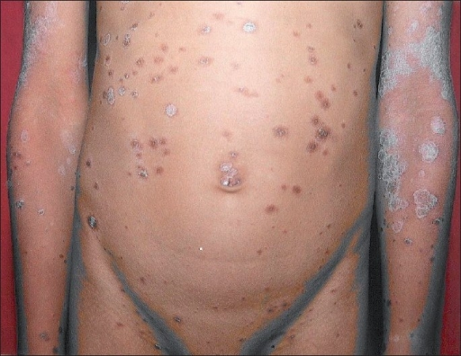
The condition usually starts with a primary plaque which, after one week, evolves into a widespread eruption of smaller skin lesions.
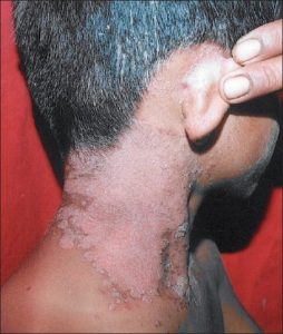
- Image: “Sudden appearance of scaly sharply demarcated erythematous lesions on side of neck in the next week.” by Zawar V. License: CC BY 2.0
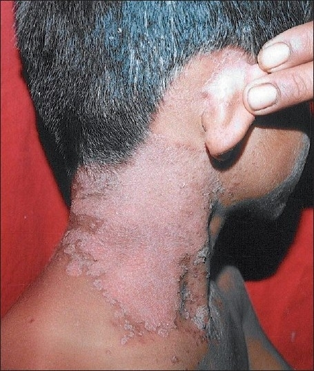

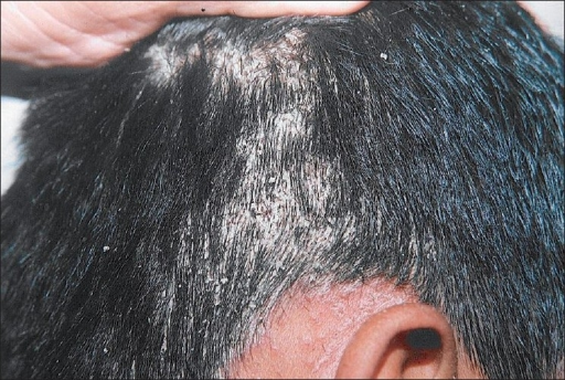
- Image: “Thick, adherent “asbestos-like” scales with buried tufts of hairs on the scalp.” by Zawar V. License: CC BY 2.0
Several studies have shown an increased incidence of atopy disorders, such as food allergies, atopic dermatitis and acne vulgaris in children with PR, a finding in favor of an immune-based mechanism of pathogenesis.
Clinical Presentation of Pityriasis Rosea in Children
Patients with PR usually do not have a recent positive travel history, or history of similar skin eruptions in other family members. Recent medication use should be excluded as many medications have been associated with a PR-like skin rash as an adverse reaction.
A recent history of an upper respiratory tract infection can be elicited in a significant number of patients. Patients can also describe a prodromal phase where they feel tired and can have non-specific symptoms. The most common symptoms in the prodromal phase include headaches, nausea, fever and joint pain.
The first sign of the disease is the eruption of a single patch that is salmon-colored and is well-demarcated. After one to two weeks, a generalized exanthema appears. The new macules are usually bilateral and symmetric and are oriented with the skin cleavage lines. Once this generalized skin rash occurs, it typically resolves within 6-8 weeks. The herald patch may not always be present. Secondary eruptions usually occur on distal parts of elbows and knees, and face.
In contrast to drug-related eruptions, PR skin eruptions are usually mildly pruritic and can be non-pruritic in up to one-quarter of the cases.
Diagnostic Workup for Pityriasis Rosea in Children
The diagnosis of PR is largely a clinical one with little room for laboratory tests. In a very small number of cases, patients might have elevated erythrocyte sedimentation rate or an increased total serum protein level or albumin level.
PR can be confused with secondary syphilis and a rapid plasma regains test should be performed to exclude the latter. When the child is too young to be expected to be involved in sexual activities and there are no alarming signs of possible sexual abuse, there is usually no need for sophisticated tests to exclude syphilis.
Differential Diagnosis
Pityriasis rosea has to be differentiated from other look alike conditions like:
- Tineacorporis- these skin lesions may look like herald patch but here there are few lesions as compared to pityriasisrosea, and secondary skin lesions do not follow. positive fungus culture confirms the diagnosis.
- GuttatePsoriasis- eruption follow upper respiratory tract infection. Skin biopsy confirms diagnosis.
- Syphillis- Patients with pityriasis like rash with history of risk factors for sexually transmitted disease, syphilis should be suspected until proved otherwise. Herald patch is absent in syphilitic rash.
Treatment of Pityriasis Rosea in Children
It is essential to confirm the diagnosis of PR and exclude any other causes of PR-like skin eruptions because the treatment of PR is largely supportive and symptomatic. Children who complain of pruritus might benefit from topical application of zinc oxide and calamine lotion.
Patients with severe and wide-spread PR might need topical or oral steroids. The use of steroids in PR is still controversial because there is no definitive proof that the condition is truly immune-mediated. Ultraviolet radiation therapy should be limited to cases of severe PR because of the increased risk of post-inflammatory pigmentation after treatment.
Patients with moderately pruritic PR, especially at night-time, should receive a sedative antihistamine at bedtime. These antihistamines help with the pruritus and can also help the child fall into sleep.
The role of antibiotic therapy in children with PR is not existent as several studies have pointed out that antibiotics add no benefits to the patient. Acyclovir has been found to result in significant symptomatic improvement of PR at 1 week, 2 weeks and 6 weeks after the onset of the widespread rash, but the total duration of the illness was not affected.
Prognosis of Pityriasis Rosea
The disease is self-limiting, may resolve within 6-8 weeks, and can last for 3-4 months. Post-inflammatory hypopigmentation or hyperpigmentation may occur. Eruptions do not leave any scar behind.


Comentários
Enviar um comentário