Imaging in Early Pregnancy
Table of Contents
- Common Causes of Acute Abdominal Pain in Early Pregnancy
- Ultrasonography in Early Pregnancy
- X-Ray in Early Pregnancy
- Magnetic Resonance Imaging in Early Pregnancy
- Specific Considerations Related to Common Causes of Pelvic Pain in Pregnant Women
- Obstetric and Gynecologic Causes of Acute Pelvic Pain in Pregnancy
- References
Common Causes of Acute Abdominal Pain in Early Pregnancy
Pregnancy is associated with several problems that may present with complains such as abdominal pain, fundal height that is greater than gestational age and exaggeration of all the irritations of pregnancy.
The most common presentation of first trimester pathology is acute abdominal pain that results from one of two types of causes.
The first type are disorders unrelated to pregnancy, such as:
- appendicitis
- a ureteric stone obstruction
- rupture of an ovarian cyst
- an adnexal torsion
- a degenerating myoma.
In the second group fall disorders related to pregnancy, including:
- placental abruption
- molar pregnancy
- miscarriages.
Preexisting diseases that increase the risk and complicate the problems of pregnancy are:
- diabetes
- hypertension.
Based on the above findings, the diagnosis and identification of the etiology of lower abdominal or pelvic pain in the pregnant woman can be challenging. Thus, routine blood workup for acute abdomen in pregnant women is challenging due to :
- The physiologic alterations induced by normal pregnancy.
- The growing uterus which can alter the anatomic location of the different pelvic organs. This makes it difficult to make a conclusive diagnosis based on the performed investigations.
- The available imaging modalities, such as X-rays, posing a great deal of harm to the patient. Therefore, one should exercise caution when deciding on the preferred modality.
In pregnancy, safe and sophisticated imaging modalities are needed for the diagnosis of acute abdomen and other first trimester conditions. These choices include ultrasounds and magnetic resonance imaging techniques.
Ultrasonography in Early Pregnancy
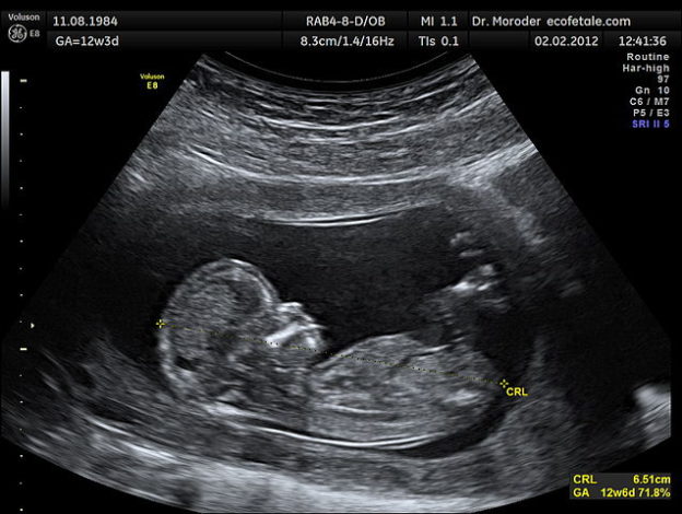
Ultrasonography remains the imaging modality of choice for the identification of various pathologies and their etiologies in the first trimester of pregnancy. Ultrasonography is known to be safe in early pregnancy and during organogenesis. The sensitivity and specificity of ultrasonography in the evaluation of the maternal pancreas, kidneys and gallbladder are excellent. During the first trimester, the uterus is not large enough to make other pelvic or abdominal organs obscure. Therefore, ultrasound is important for:
- Identification and assessment of the cause of abdominal pain that may not be related to pregnancy, such as acute appendicitis. Ultrasonography is commonly used in pregnant women suspected to have appendicitis. Unfortunately, the visualization of the appendix with ultrasonography can be difficult. Therefore, the use of ultrasonography for the evaluation of a patient with acute appendicitis is usually indicated for the exclusion of other obstetric or gynecological causes of pelvic pain rather than confirming the diagnosis of appendicitis.
- Identification and assessment of the cause of abdominal pain that may be related to pregnancy, such as ectopic pregnancy, molar pregnancy, threatened abortion and incomplete abortion. During the third trimester ultrasound is used to assess the position of the placenta, fetal cardiac activity and breathing movements.
- Diagnosis and dating of the pregnancy when the last normal menstrual period is uncertain.
- Assessment of fetal development and identification of early congenital abnormalities. Ultrasonography is also used in early pregnancy for calculation of the gestational age, and the confirmation of fetal viability. During the second and third trimesters, ultrasound is used to exclude congenital anomalies, assess the amniotic fluid volume, asses the placenta and evaluate the overall well-being of the fetus.
- To guide therapeutic procedures such as chorionic villus sampling, cervical cerclage and amniocentesis.
- Determination of multiple gestation, its chorionicity and amnionicity.
X-Ray in Early Pregnancy
Plain radiography or computed tomography (CT)
Plain radiography or computed tomography (CT) scans might be needed in early pregnancy for the evaluation of certain conditions. It should be noted that with conventional x-ray machines, the radiation dose from a single exposure is too low to be associated with any fetal anomalies or fetal loss.
Pelvic radiography
Pelvic radiography might be used for the evaluation of urolithiasis in a pregnant woman, but ultrasonography can provide excellent visualization of the stone in experienced hands without exposure to ionizing radiation.
Abdominal radiography
Abdominal radiography might be indicated in a pregnant woman suspected to have small bowel obstruction. Again, a single x-ray exposure is very unlikely to cause any harm to the fetus.
CT scans of the lower abdomen or pelvis
CT scans of the lower abdomen or pelvis might be warranted for the evaluation of certain conditions, i.e., acute pancreatitis or appendicitis. While the radiation dose of a single CT scan of the pelvis is around 10 mGy, it should still be used with caution in pregnant women. The risk of fetal anomalies or fetal loss after a single pelvic or abdominal CT scan is almost zero; however, there appears to be an increased risk of childhood cancer in the offspring who were exposed to ionizing radiation during pregnancy.
Fetal anomalies: Radiation exposure of more than 200 mGy (about 20 pelvic CT scans) during the first 2–8 weeks of gestation is associated with fetal anomalies.
Mental retardation: This is common after an exposure that is greater than 500 mGy at 8–12 weeks of gestation and an exposure that is greater than 250 mGy at 16–25 weeks of development.
Clearly, these numbers are very comforting to the radiologist as they clearly show that a single CT exposure during early pregnancy will not cause fetal anomalies or loss. Despite this, ionizing radiation remains a second-line imaging modality in the diagnostic workup of acute abdomen in pregnant patients.
Magnetic Resonance Imaging in Early Pregnancy
The sensitivity of magnetic resonance imaging for acute appendicitis in pregnant women is very high. Magnetic resonance imaging is also superior to other imaging modalities in the evaluation of the fetus and for excluding fetal anomalies. MRI during early pregnancy is most likely harmless; however, contrast agents are rarely administered unless they are necessary to answer the doctor’s questions. Ultrasonography remains the imaging modality of choice for the diagnostic workup of acute appendicitis in pregnant patients.
Specific Considerations Related to Common Causes of Pelvic Pain in Pregnant Women
Acute Appendicitis in Pregnant Women
Symptoms
Nausea is present in all cases of appendicitis in pregnant women. Vomiting is present only in two thirds. While anorexia is present in all non-pregnant patients with acute appendicitis, it is only present in one third of pregnant women who have acute appendicitis. Tenderness is usually well localized in the right lower quadrant in the first trimester.
White blood cell counts can be as high as 15,000/microliter in a healthy pregnant woman. Therefore, white blood cell counts are not useful in the evaluation of acute appendicitis in pregnant patients.
Diagnosis
Ultrasonography can be used to exclude other causes of pelvic pain such as adnexal torsion, ruptured ovarian cyst or ruptured ectopic pregnancy. Ultrasonography might also show peri-appendicular edema and fluid collection, findings suggestive of acute appendicitis. Patients that are equivocal should undergo either a computed tomography scan of the pelvis or a magnetic resonance imaging study. Both imaging modalities provide an almost 100 % sensitivity and specificity for acute appendicitis.
Bowel Obstruction in Pregnant Women
Abdominal pain is present in most patients. Radiation of the pain to the flanks is more common in pregnant women compared to non-pregnant women with acute bowel obstruction.
Constipation is common during pregnancy, therefore, one should ask specifically about any differences between their usual constipation and the current bowel movement habits.
Abdominal tenderness with high-pitched bowel sounds is not seen in pregnant women with acute bowel obstruction. Bowel sounds are usually normal on auscultation in contrast to the absence of bowel sounds in non-pregnant patients with acute bowel obstruction.
In acute bowel obstruction, the imaging modality of choice is an upright plain radiograph of the abdomen. The radiation dose is minimal from a single radiograph.
Urolithiasis in Pregnancy
Flank pain is present in almost all cases of urolithiasis. Nausea, vomiting, dysuria, fever and hematuria are also common findings. In one quarter of the patients, history of a prior episode of ureteric or renal colic is present. Costovertebral angle tenderness is present in all cases.
The imaging modality of choice in the pregnant patient is ultrasonography. Ultrasonography is used to check for signs suggestive of obstruction rather than the actual visualization of the stone itself. Right-sided ureteric and calyces’ dilation can be seen in a normal pregnancy.
Obstetric and Gynecologic Causes of Acute Pelvic Pain in Pregnancy
Ovarian Cyst
A ruptured ovarian cyst presents with a history of mild trauma. Pelvic pain is common. The patient might be in shock if severe hemorrhage occurs. Ultrasonography is the imaging modality of choice. It can show free fluid in the cul-de-sac in case of a ruptured ovarian cyst.
Adnexal torsion
Adnexal torsion also presents with acute, severe, colicky, unilateral, lower pelvic pain. Nausea and vomiting are found in two thirds of the patients. A tender adnexal mass is found in almost all patients.
Ultrasonography with color Doppler is useful in confirming the diagnosis and assessing ovarian blood flow to the central ovarian parenchyma respectively. Diagnostic laparoscopy is indicated in difficult equivocal cases.
Degenerating myoma
A degenerating myoma presents with very well localized abdominal or pelvic acute pain, tenderness and vomiting. Ultrasonography can be used to confirm the diagnosis. The probe should be placed directly on the painful area.
Placental abruption
Placental abruption can happen in hypertensive patients, those who use cocaine and cigarette smokers. Vaginal bleeding, pelvic and back pain, hypertonic uterus and non-reassuring fetal heart rate are the most common findings in placental abruption. Ultrasonography can be used. If negative, a magnetic resonance imaging study is indicated. Ultrasonography can detect only 25 % of the cases of placental abruption.


Comentários
Enviar um comentário