Anatomy of the Forearm
Table of Contents
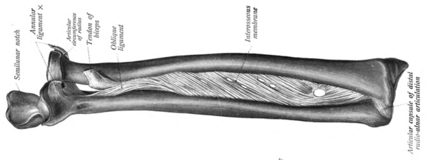
Image: “Diagram showing radius and ulna, the interosseous membrane between them as well as the paoximal and distal radioulnar joints.” by Dr. Johannes Sobotta – Sobotta’s Atlas and Text-book of Human Anatomy 1909. License: Public Domain
Gross Anatomy of the Forearm
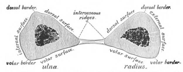
The radius and ulna are joined by a fibrous tissue called the interosseous membrane that helps the bones function together during pronation and supination as well as dividing the forearm into anterior and posterior compartments.
Image: “Diagram showing interosseous membrane expanding between radius and ulna.” by Dr. Johannes Sobotta – Sobotta’s Atlas and Text-book of Human Anatomy 1909. License: Public Domain
Radius
The proximal part of the radius articulates with the ulna. It is narrow and cylindrical and is made up of the neck and radial tuberosity.
The distal part of the radius contains articulation sites for the carpal bones, the scaphoid and lunate.
Cross section: triangular/prism shape.
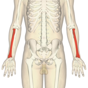
Image: “Diagram identifying radius of both forearms.” by Anatomography – en:Anatomography (setting page of this image). License: CC BY-SA 2.1 JP
Three borders:
- Anterior or volar: Arises from the radial tuberosity and makes an oblique line of the radius as it crosses from medial to lateral. It extends from the tuberosity to the styloid process. Towards the end of the anterior surface, a tubercle with the tendon of brachioradialis muscle is found.
- The following muscles have origins on the volar border: flexor digitorium superficialis muscle, flexor pollicis longus muscle.
- The following muscles have insertions on the volar border: supinator muscle, pronator quadratus muscle as well as the dorsal carpal ligament.
- Posterior or dorsal: most visible on the middle third of the bone. This border also ends at the posterior part of the styloid process.
- Interosseous: sharp border which is continuous with the interosseous membrane. It ends at the ulnar notch.
Three surfaces:
- Anterior or volar: smooth, concave and makes up the upper ¾ of the radius
- The following have origin on the volar surface: flexor pollicis longus muscle, volar radiocarpal ligament
- The following have insertion on the volar surface: pronator quadratus
- Posterior or dorsal: upper and middle thirds are concave whereas the lower 1/3 is convex.
- The following have the origin in the upper 1/3: supinator
- The following have the origin in the middle 1/3: abductor pollicis longus, extensor pollicis brevis
- Tendons of various muscles take origin in the lower 1/3
- Lateral: entirely convex, hence known as the “convexity of the radius”. Contains the oval, rough area that serves as insertion sites for the pronator teres and supinator muscles.
Ulna
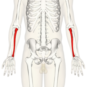
Image: “Diagram identifying the ulna of both forearms.” by Anatomography – en:Anatomography (setting page of this image). License: CC BY-SA 2.1 JP
The proximal part of the ulna is large whereas the distal part is narrow. The styloid process is found on the distal end of the ulna. The olecranon process of the ulna articulates with the olecranon fossa of the humerus and forms a hinge joint at the elbow. This articulation also prevents hyperflexion at the elbow.
Another structure, called the radial notch, accommodates the head of the radius on the ulna.
Like the radius, the ulna also consists of three borders; anterior, posterior and interosseous, the posterior border being the sharpest and distinct and interosseous border being continuous with the interosseous membrane that attaches the ulna and radius together.
Three surfaces:
- Anterior or volar surface: initially smooth but becoming rough in texture at the distal end due to insertion of pronator quadratus muscle.
- Medial surface
- Posterior surface
The following muscles have origins on the ulna:
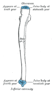
Image: “Illustration showing proximal and distal ends of the ulna.” byHenry Vandyke Carter, Henry Gray (1918) Anatomy of the Human Body. Bartleby.com: Gray’s Anatomy, Plate 215. License: Public Domain
- Pronator teres muscle
- Supinator muscle
- Flexor carpi ulnaris muscle
- Flexor digitorium superficialis muscle
- Extensor carpi ulnaris muscle
- Extensor pollicis longus muscle
- Abductor pollicis longus muscle
- Flexor digitorium profundus muscle
The following muscles have insertions on the ulna:
- Brachialis muscle
- Tricep brachii muscle
Muscles found in the forearm bring about the following movements:
Flexion of the wrist and digits of hands; that is, decreasing the angle between the structures and their proximal segments.
Extension of wrist and digits of hands; that is, increasing the angle between the structures and their proximal segments.
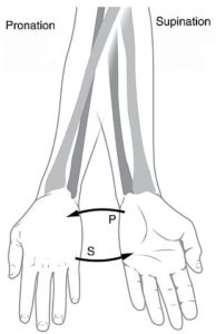
Pronation of the forearm and hand is when they are turned inwards; that is, the palm is facing the ground.
Supination of the forearm and hand is when they are turned outwards; that is, the palm is facing the sky.
Forearm in cross-section:
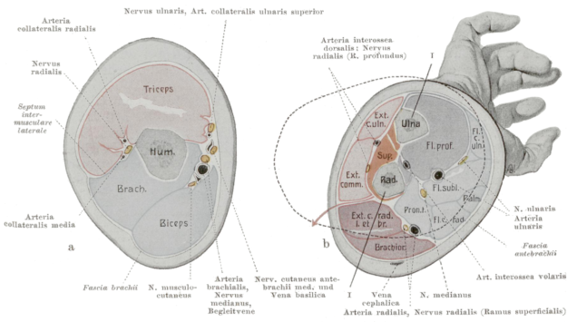
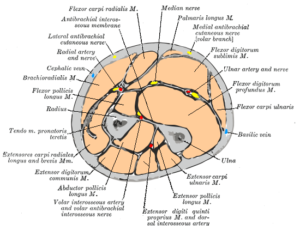
The cross-section of the forearm shows two fascial compartments. The following structures can be viewed from a cross-section of the forearm:
- Bones: radius and ulna
- Nerves: median nerve, ulnar nerve, lateral and medial cutaneous nerve of the forearm, radial nerve
- Arteries: radial and ulnar arteries
- Veins: cephalic and basilic veins
- Muscles: Extensor carpi radialis longus, flexor carpi radialis, flexor digitorium superficialis, flexor carpi ulnaris, flexor digitorium profundus, flexor pollicis longus, extensor digitorium, extensor carpi ulnaris, extensor pollicis ulnaris, extensor pollicis longus.
Fascial compartments comprise of groups of muscles and nerves with similar functions contained within the same fascia. The forearm is made up of two compartments; anterior and posterior compartments.
The structures dividing the forearm into these compartments are as follows:
- Interosseous membrane
- Lateral intermuscular septum that extends from the anterior border of the radius to the deep fascia around the upper limb
- Deep fascia attachment along the posterior border of the ulnaImage: “Diagram showing the cross-section of the upper arm (left) and forearm (right).” by Braus, Hermann – Anatomie des Menschen: ein Lehrbuch für Studierende und Ärzte. License: Public Domain
Anterior Compartment of the Forearm
The anterior compartment of the forearm is also known as the flexion compartment because it works to flex the forearm and wrist, as well as pronate the hand.
Image: “Diagram showing cross-section of anterior compartment of the forearm.” by Henry Vandyke Carter, Henry Gray (1918) Anatomy of the Human Body. Bartleby.com: Gray’s Anatomy, Plate 417. License: Public Domain
There are three layers of muscles in the anterior compartment:
- Superficial
- Intermediate
- Deep
Muscles, along with their functions and attachments, belonging to the superficial layer of the anterior compartment, include the following:
- Flexor carpi ulnaris – originates from the humeroulnar head and inserts in the pisiform bone. Works to flex and adduct the wrist.
- Palmaris longus – originates from the medial epicondyle of the humerus and inserts in the palmar aponeurosis of the hands. Works to flex the wrist joint.
- Flexor carpi radialis – also originates from the medial epicondyle of the humerus and inserts in the base of 2nd and 3rd metacarpals. Works to flex and abduct the wrist.
- Pronator teres – originates from the medial epicondyle of the humerus and inserts in the lateral surface of the radius. Works to pronate the forearm.
The only muscle belonging to the intermediate layer of the anterior compartment is the flexor digitorium superficialis. It emerges from the humeroulnar head and inserts as four separate tendons on the palmar surfaces of the middle phalanges of the index, middle, ring and little fingers. It works to flex the middle interphalangeal joints of the fingers it attaches to.
Muscles, along with their functions and attachments, belonging to the deep layer of the anterior compartment, include the following:
- Flexor digitorium profundus – originates from the anteromedial surface of ulna and inserts as four different tendons on the palmar surface of the distal phalanges of the index, middle, ring and little fingers. Works to flex the middle interphalangeal joints of the fingers it attaches to.
- Flexor pollicis longus – originates from the anterior surface of the radius and inserts palmar surface of the distal phalanges of the thumbs. Works to flex the interphalangeal joint of the thumb.
- Pronator quadratus – originates from the anterior surface of distal ulna and inserts in the anterior surface of the distal radius. Works to pronate the forearm.
Arterial Supply of the Anterior Compartment of the Forearm
Two main arteries of the forearm run through the anterior compartment. They are the division of the brachial artery from the upper arm: the radial and ulnar arteries.
The radial artery emerges from the brachial artery at the neck of the radius. Its clinical significance lies in the measurement of the radial pulse by locating the tendon of the flexor carpi radialis muscle in the distal forearm.
The radial artery gives rise to the following branches:
- Radial recurrent artery – anastomoses around the elbow
- Palmar carpal branch – anastomoses in the hand to supply the carpal bones
- Superficial palmar branch – anastomoses with the superficial arch of the ulnar artery.
The ulnar artery passes down the medial side of the forearm and is not easily palpable because it lies deep to the flexor carpi radialis muscle in the distal forearm.
The ulnar artery gives rise to the following branches:
- Ulnar recurrent artery – anastomoses around the elbow
- Common interosseous artery – gives rise to anterior and posterior interosseous artery
- Dorsal and palmar carpal branches – supply the carpal bones.
All the muscles of this compartment are innervated by the median nerve, an exception being flexor carpi ulnaris and flexor digitorium profundus’ medial half, which are innervated by the ulnar nerve.
Posterior Compartment of the Forearm
Image: “Diagram showing the superficial muscles of the posterior compartment of the forearm.” by Henry Vandyke Carter, Henry Gray (1918) Anatomy of the Human Body. Bartleby.com: Gray’s Anatomy, Plate 418. License: Public Domain
The posterior compartment of the forearm is also known as the extension compartment because it causes the extension of the forearm, wrist, and fingers as well as their supination.
Muscles of the posterior compartment are divided into superficial and deep layers.
The muscles, along with their functions, innervations, and attachments, belonging to the superficial layer of the posterior compartment, include the following:
- Brachioradialis – originates from the proximal lateral supraepicondylar ridge of the humerus and inserts on the lateral surface of the distal end of the radius. It works to help flex the elbow while the forearm is mid-pronated. Innervation: radial nerve (C5, C6)
- Extensor carpi radialis longus – also originates from the lateral supraepicondylar ridge of the humerus and inserts on the dorsal surface of the 2nd metacarpal. It works to extend and abduct the wrist. Innervation: radial nerve (C6, C7)
- Extensor carpi radialis brevis – It originates from the lateral epicondyle of the humerus, inserts in the dorsal surface of the 2nd and 3rd metacarpal. It also works to extend and abduct the wrist. Innervation: deep branches of radial nerve (C7, C8)
- Extensor digitorium – also originates from the lateral epicondyle of humerus and inserts as four separate tendons on the dorsal parts of the bases of middle and distal phalanges of the index, middle, ring and little fingers. It works to extend the fingers it is attached to. Innervation: posterior interosseous nerve (C7, C8)
- Extensor digiti minimi – also originates from the lateral epicondyle of the humerus and inserts in the hood of the little finger. It works to extend the little finger. Innervation: posterior interosseous nerve (C7, C8)
- Extensor carpi ulnaris: also originates from the lateral epicondyle of the humerus and inserts in the base of the medial side of the 5th metacarpal. It works to extend and adduct the wrist. Innervation: posterior interosseous nerve (C7, C8)
- Anconeus: originates from the lateral epicondyle and inserts in the olecranon. Works to help extend the elbow and abduct the ulna during pronation. Innervation: radial nerve (C6, C7, C8)
Muscles, along with their functions, innervations, and attachments, belonging to the deep layer of the posterior compartment, include the following:
- Supinator: originates from a superficial (lateral epicondyle of the humerus) and deep (supinator crest of ulna) part and inserts in the lateral surface of the radius. It works to supinate the forearm. Innervation: posterior interosseous nerve (C6, C7)
- Abductor pollicis longus: originates from the posterior surface of both radius and ulna and inserts on the lateral side of the 1st metacarpal. It works to abduct the thumb. Innervation: posterior interosseous nerve (C6, C7)
- Extensor pollicis brevis: originates from the posterior surface of the radius and inserts in the dorsal surface of the base of the distal phalanx of the thumb. It works to extend the thumb. Innervation: posterior interosseous nerve (C6, C7)
- Extensor pollicis longus: originates from the posterior surface of the ulna and inserts on the dorsal surface of the base of the distal phalanx of the thumb. It works to extend the thumb. Innervation: posterior interosseous nerve (C6, C7)
- Extensor indicis: originates from the posterior surface of the ulna and inserts in the extensor hood of the index finger. It works to extend the middle finger. Innervation: posterior interosseous nerve (C6, C7).
The anterior and posterior interosseous arteries, along with the radial artery, are the main arterial supply of the posterior compartment of the forearm.
The main nerve of the posterior compartment is the radial nerve, which gives rise to the posterior interosseous nerve along with its superficial and deep branches.

Comentários
Enviar um comentário