Cell Division: Meiosis
Table of Contents
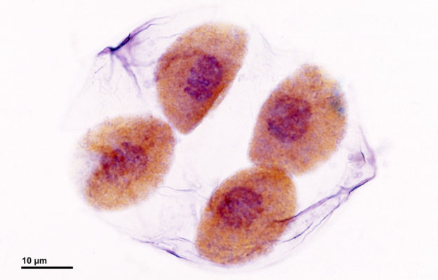
Image: “Meiosis: Dividing pollen mother cells (at the end of meiotic division) – Lilium plant. Optical microscopy technique: Bright field. Magnification: 3000x.” by Doc. RNDr. Josef Reischig, CSc. – Author’s archive. License: CC BY-SA 3.0
Overview of Meiosis
Meiosis is the process by which eukaryotic cells produce daughter cells that are different from the mother cell and from each other. This is possible because in the whole process of meiosis, some regions of DNA in the chromosome have been shuffled through crossing over.
Independent assortment also occurs because of shuffling of whole chromosomes. The end product of meiosis are daughter cells with half the number of chromosomes as the original one. These processes occur in the ovaries and testes producing the daughter cells which are called gametes.
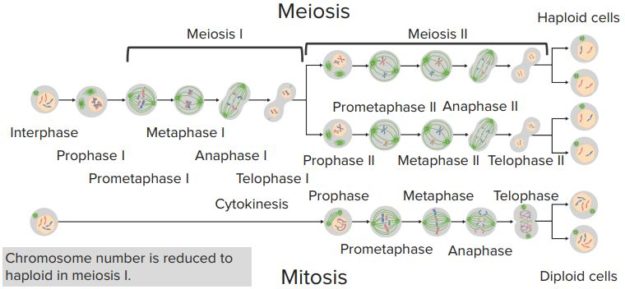
Meiosis consists of two rounds of cell division. These are called meiosis I and meiosis II. Each of the rounds is divided into different phases namely prophase, metaphase, anaphase, telophase and cytokinesis.
Each of these phases contributes to the production of unique daughter cells. Distinguishing features of meiosis include synapsis pairing where homologous chromosomes pair up with each other. During the synapsis, crossing over between the two chromosomes results to genetic recombination.
Meiosis I
Prophase I
The first part of meiosis I is prophase I. The prophase stage is a long process that can be subdivided into five stages. The 1st stage is the leptotene stage, where the chromosomes start to condense.
The 2nd stage is the zygotene stage where homologous chromosomes align directly opposite each other and are held together at several points along their length. This is when synapsis between homologous chromosomes occurs.
The 3rd stage is the pachytene stage, where the chromosomes condense and become shorter and thicker and each pair of the homologous chromosome becomes tightly coiled.
The 4th stage is the diplotene stage, where crossing over occurs and is visible because of the chiasmata – the point of crossing over. In this stage, the homologous recombinant chromosomes begin to separate but remain attached at the chiasmata.
The last stage is the diakinesis where separation of the homologous chromosome pairs proceeds as the chromosomes become fully condensed.
Metaphase I
After prophase I, metaphase I proceeds. This is when the nuclear membrane disassembles. Chromosomes align themselves on the equatorial plane and become attached to spindle fiberssimilar during metaphase of mitosis. These spindle fibers push the chromosome pairs to stay in the middle of the cell.
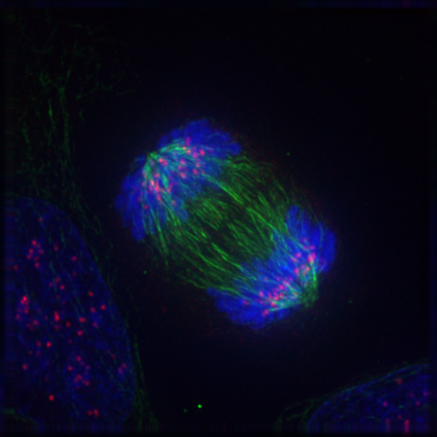
Image: “Metaphase. Chromosomes lined up on the metaphase plate. Two views rotated 90°. Produced using anti-dsDNA antibodies on HEp-20-10 cells with a FITC conjugate.” by Simon Caulton. License: CC BY-SA 3.0
Anaphase I
Anaphase I then proceeds and is characterized by shortening of the spindle fibers. The shortening of the spindle fibers results in separation of the chromosomes in the pairs.
Telophase I and cytokinesis
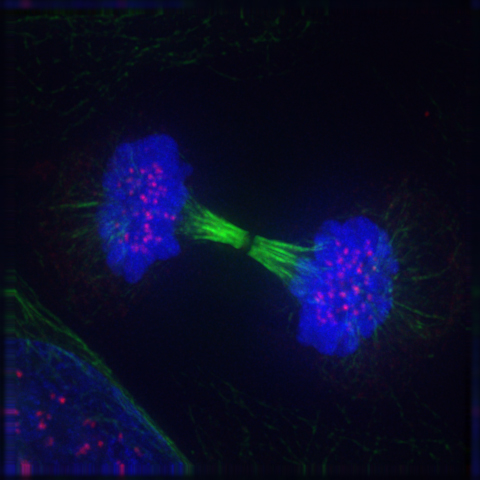
Image: “Reappearance of the nuclear membrane and nucleolus: the telophase” by Roy van Heesbeen. License: Public Domain
The spindle fibers then disassemble during Telophase I. Chromosomes become less condensed here and nuclear envelope may start to reform. Cytokinesis then proceeds dividing the cell into two daughter cells, which contain only one set of chromosomes and are considered haploid.
Meiosis II
Meiosis II proceeds in a process similar to an ordinary mitotic division. Each chromosome, which exists as a pair of chromatids, align along the center of the cell, then split to produce two new daughter gametes known as ova or spermatids. Like meiosis I, meiosis II is divided into stages namely prophase II, metaphase II, anaphase II, telophase II and cytokinesis.
If the nuclear envelope is reformed during meiosis I, it will again disassemble during prophase II. Spindle fibers again form and chromosomes start to condense. The spindle fibers again push the chromosomes to the middle of the cell as it elongates during metaphase II.
The sister chromatids are separated when the spindle fibers shorten during anaphase II. The chromosomes then become less condensed and the spindle fibers disassemble during telophase II. It is also during this stage that the nuclear envelope reforms. Cytokinesis then divides the cell into two daughter cells, that are haploid.
Meiosis in Humans
The human karyotype contains normally 23 pairs of chromosomes. Body cells are diploid cells containing 46 chromosomes, which includes already the maternal and the paternal chromosomes. This complete set of chromosomes contains 22 pairs of autosomes and a pair of allosome.
Autosomes are the chromosomes that determine which proteins are made in the body, while allosomes are the chromosomes involved in the determination of sex. Meiosis is important in the production of the haploid cells containing the gametes. The spermatocytes and the oocytes are the two types of haploid cells produced during meiosis.
During sexual reproduction, these two haploid cells combine to produce the diploid cell containing the chromosomes both from the mother and the father. The spermatocytes are produced in the process called spermatogenesis while the oocyte is produced in the process called oogenesis.
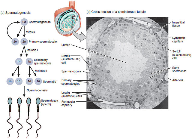
Spermatogenesis
Spermatogenesis occurs in the seminiferous tubules of the testis. The whole process can be divided into three stages namely spermatogonial stage, meiotic stage and spermiogenesis stage.
In the spermatogonial stage, mitotic clonal expansion occurs. The type A spermatogonia which arose from the primordial germ cell divide to maintain stem cell pool population. Some of the type A spermatogonia return or stay at the resting pool while some proliferate and undergo differentiation.
In the process during the spermatogonial stage, the type A spermatogonia may be converted to type B spermatogonia, which are the precursor cell for spermatocytes. The type B spermatogonia then enter the preleptotenes stage of the meiotic process and are now called primary spermatocyte.
The primary spermatocytes undergo the two stages of the meiotic process. The resulting cell after the whole meiotic division are the haploid daughter cells which are called round spermatids.
The round spermatids then enter the spermiogenesis stage. Here, spermatids undergo a number of nuclear and cytoplasmic changes leading to the formation of the spermatozoa.
These changes include formation of the acrosome which is a modified lysosome. The nucleusbecomes condensed and is moved to the periphery of the cell. Microtubules then form producing a flagella. In this process, a large part of the cytoplasm is removed as a residual body. The Sertoli cells phagocytose these bodies. The spermatozoa produced are then stored to mature before released into the epididymis.
Oogenesis
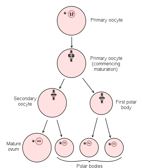
Image: “Diagram showing the reduction in number of the chromosomes in the process of maturation of the ovum. (In mammals, the first polar body normally disintegrates before dividing, so only two polar bodies are produced.)” by Henry Vandyke Carter – Henry Gray (1918). License: Public Domain
Oogenesis, unlike spermatogenesis, involves certain gaps in its process. Before birth, the oogonium, which also arose from the germs cells, undergoes mitosis to produce the primary oocyte. It starts to undergo meiosis I but the process is halted at the prophase I.
After puberty, the primary oocytes complete meiosis Iforming a secondary oocyte and a first polar body. The secondary oocyte then begins meiosis II and is ovulated. The secondary oocyte is arrested in metaphase II.
When fertilization occurs between the secondary oocyte and a sperm cell, meiosis II proceeds and is completed when the sperm cell completely penetrates the secondary oocyte. A second polar body and the ovum is produced after meiosis II. When the nuclei of the sperm cell and the ovum unite, a diploid zygote is produced.
Meiosis vs. Mitosis
One major difference between these two processes is the number of steps needed to complete cell division.
Mitosis involves a single division producing two daughter cells that are similar to the parent cell.
Meiosis occurs as a two cell division process which produces 4 daughter cells that are completely distinct from each other and from the parent cells. The final product of meiosis has half the number of chromosomes compared to the parent cell while mitosis produces daughter cells with the same number of chromosomes.
Mitosis occur in somatic cells and during early cell division in gamete formation while meiosis occurs only at the final division of gamete maturation.
“Comparing Mitosis & Meisosis” Image created by Lecturio
Review Questions
The correct answers can be found below the references.
1. In which stage of the prophase I do the homologous chromosomes start to separate and the crossing over becomes more visible because of the chiasmata?
- Leptotene
- Diplotene
- Pachytene
- Zygotene
2. Which of the following statements correctly describes the product of complete meiotic cell division?
- 2 diploid daughter cells that equal one another and the parent cell
- 2 diploid daughter cells that are the same as the parent cell
- 4 haploid daughter cells that differ from one another and the parent cell
- 4 haploid daughter cells that equal one another but differ from the parent cell.


Comentários
Enviar um comentário