Rhombencephalon – Anatomy and Function of the Hindbrain
Table of Contents

Embryonic Development of the Rhombencephalon
During embryonic development, the brain, the spinal cord and the central nervous system grow from the neural tube, which itself developed from the dorsal surface ectoderm. Three primary cerebral ganglia develop from the cranial part of the neural tube.
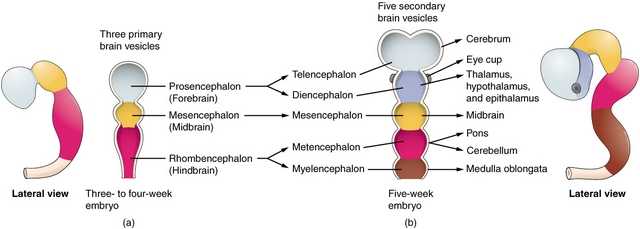
One of these cerebral ganglia becomes the rhombencephalon. The other two cerebral ganglia form the prosencephalon (forebrain) and the mesencephalon (midbrain). The cerebellum, medulla oblongata and pons proceed to grow from the rhombencephalon.
Structure of the Metencephalon
The cerebellum
Topography of the cerebellum
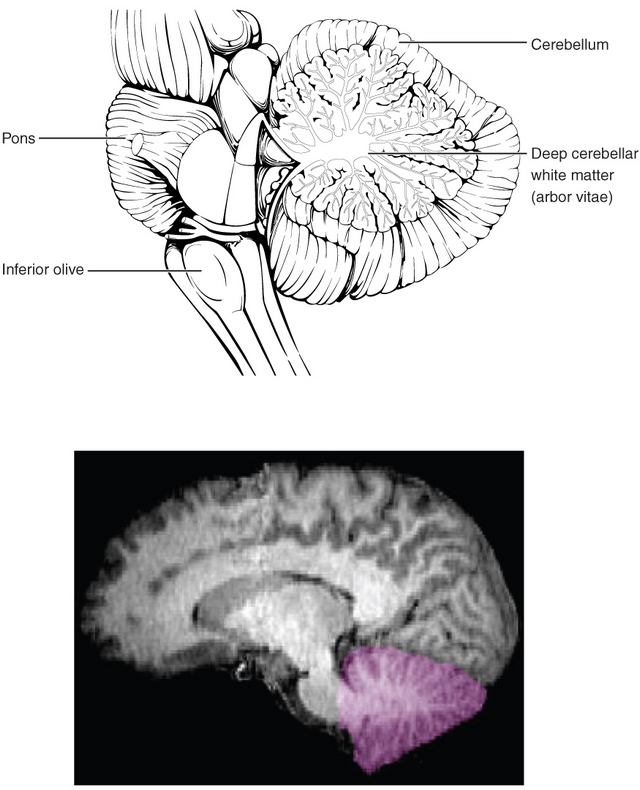
Topographically, the cerebellum is located dorsal to the medulla oblongata and the pons. The mesencephalon and medulla oblongata are connected to this via the cerebellar veil (superior and inferior medullary velum).
In addition, the cerebellum forms the roof of the fourth ventricle. The tentorium cerebelli, which forms a duplication of the dura mater, is located apical to the cerebellum.
As the dura mater consists of taut conjunctive tissue, an increase in pressure from a cerebral edema may result in impaction of the cerebellum in the tentorial incisure. This constriction is also referred to as upper impaction.
Lower impaction describes the rostral movement of the brain with impaction of the cerebellum in the foramen magnum. During lower impaction, pressure of the cerebellar tonsils on the respiratory center may be fatal.
Macroscopic structure of the cerebellum
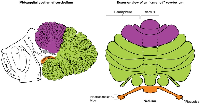
The cerebellum arises from the rhombic lips or alar plates of the metencephalon. Grossly it appears as an ovoid structure that resides in the posterior cranial fossa inferior to the tentorium cerebelli. In an anatomical specimen, the cerebellum can macroscopically be divided into the vermis (cerebellar worm) and the two hemispheres. The outer surface of the cerebellum exhibits numerous grooves (fissurae) and twists (foliae), which lead to a significant increase in the cerebellum’s surface area.
The cerebellum can also be divided into three more sections. These are the central vermis(cerebellar worm), the nodulus located in the lower part of the vermis and the flocculus located to the side of the nodulus. The nodulus and flocculus are jointly referred to as the lobus flocculonodularis.
The vermis connects the two cerebellar hemispheres, whereby each hemisphere can be divided into three lobes. These are – along with the lobus flocculonodularis – the lobus anterior and the lobus posterior.
The fissura prima is located between the two lobes. Two additional fissures in the cerebellum are the fissura horizontalis and the fissura posterolateralis, which separates the lobus posterior from the lobus flocculonodularis.
Depending on its function, the cerebellum can also be divided into three entities. These are the vestibulocerebellum, the spinocerebellum and the pontocerebellum. Their names stem from the main portion of the afferents that enter each respective entity.
The vestibulocerebellum draws most of its afferents from the vestibular system, and in its area corresponds to the lobus flocculonodularis. The spinocerebellum primarily receives its afferents from the spinal cord and consists of the cerebellar vermis and the surrounding areas. The afferents from the pontine nuclei are drawn to the pontocerebellum, which consists of the two cerebellar hemispheres.
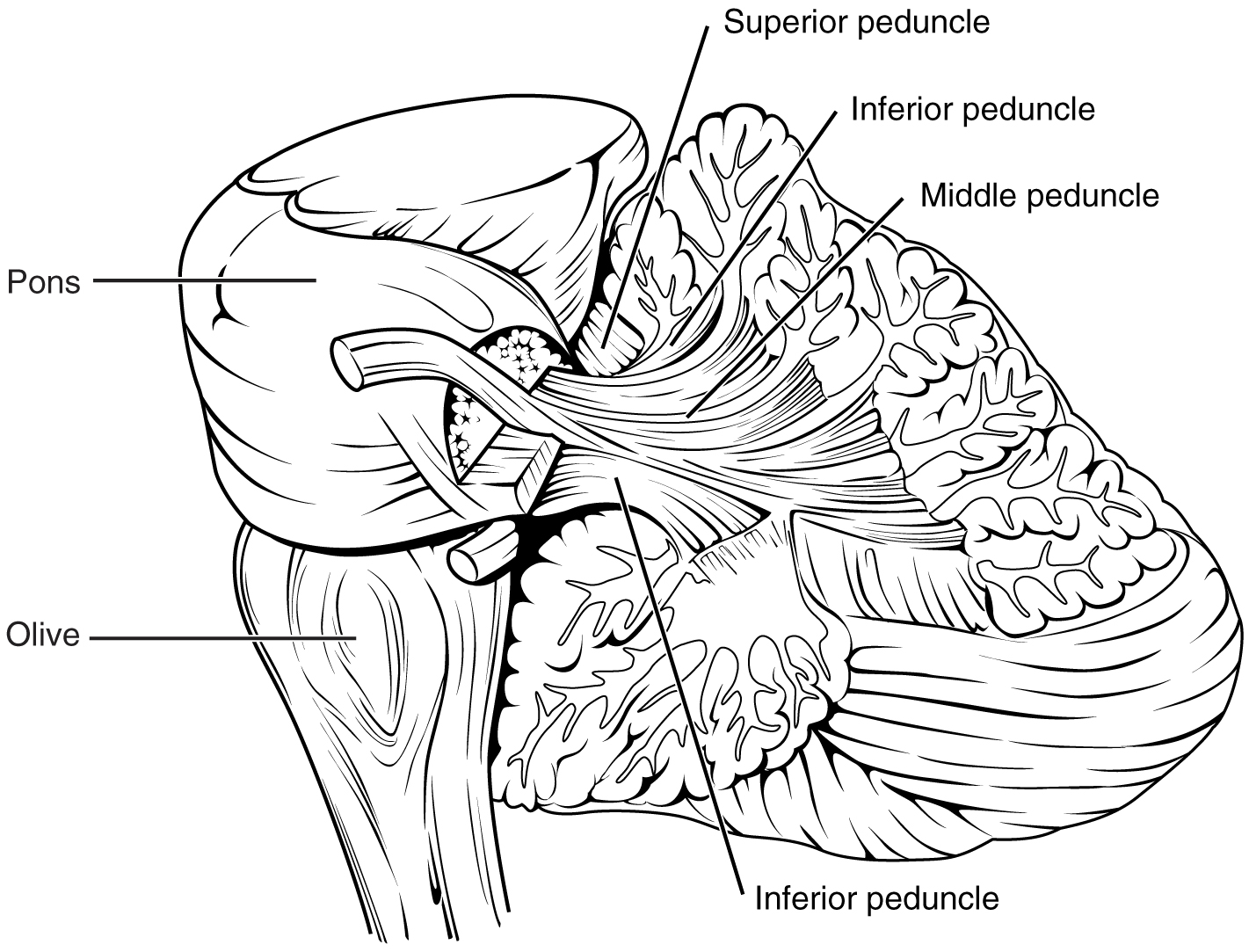
The efferents and afferents of the cerebellum pass through the three cerebellar peduncles, i.e., the pedunculus cerebellaris superior, medius and inferior.
Structure of the cerebellar cortex
The neurons of the cerebellum are located in the cerebellar cortex. Histologically, the cerebellar cortex is divided into three layers. From the inside out, there is the granular layer (stratum granulosum), the Purkinje layer (stratum purkinjese) and the molecular layer (stratum moleculare).
As the name implies, the granular layer largely consists of granule cells. These transmit their information through the stimulating transmitter glutamate. The mossy fibers, which contain all of the afferents of the cerebellum, end at the granule cells – except for afferents from the olivary bodies. The fibers extending from the olivary bodies are referred to as climbing fibers.
The Purkinje layer of cells consists of the Purkinje cells, which are the largest cells in the cerebellar cortex. Their cell bodies (somata) are located in the cerebellar hemispheres and their cell processes (axons) advance to the cerebellar nuclei (see below) and the molecular layer.
The stratum moleculare is the outermost layer of the cerebellar cortex.
Cerebellar nuclei
The cerebellar nuclei consists of four halved areas. These are the nucleus dentatus, the nucleus emboliformis, the nucleus globosus and the nucleus fastigii.
The nucleus dentatus (= toothed) is located in the center of the cerebellum. Medial to this is the nucleus emboliformis (= plug-shaped), and medial to it is nucleus globosus (= sphere-shaped). The nucleus fastigii (= gable) is the most medially located.
Afferents of the cerebellum
The cerebellum receives many afferents that pass through the three cerebellar peduncles to the cerebellar cortex. On the way, some collaterals branch out into the cerebellar nuclei.
The tractus vestibulocerebellaris, the tractus olivocerebellaris, the tractus reticulocerebellaris and the tractus spinocerebellaris posterior are among the afferents that pass through the pedunculus cerebellaris inferior.
“Vestibulocerebellum. Function and Input/Output” Image created by Lecturio
The tractus vestibulocerebellaris receives its information through the nuclei vestibulares, and this information largely ends in the area of the lobus flocculonodularis.
The fibers of the olivary bodies cross to the other side at the level of the brain stem and then pass through the tractus olivocerebellaris to the cerebellum. Thus they reach the Purkinje cells of the cerebellar cortex in the form of climbing fibers.
Proprioceptive information from the spinal cord is forwarded to the cerebellum via the tractus reticulocerebellaris.
The tractus spinocerebellaris posterior stems from the posterior horn of the spinal cord, where the nucleus dorsalis is located. The information passed through the tractus originates in the ipsilateral half of the body and reaches the granular layer in the area of the spinocerebellum as mossy fibers.
The fibrae pontocerebellaris of the nuclei pontis pass through the pedunculus cerebellaris medius and are a continuation of the corticopontine pathways. The fibers cross over to the other side before passing through the middle cerebellar peduncle and move to the cortex of the cerebellar hemispheres, i.e., the area of the pontocerebellum. The cerebellum receives information on planned movements of the cerebrum via the fibrae pontocerebellares.
The tractus spinocerebellaris anterior is an afferent of the pedunculus cerebellaris superior. It feeds information from the ipsilateral half of the body to the spinocerebellum.
Efferents of the cerebellum
The efferent pathways of the cerebellum pass from the cerebellar nuclei to the thalamus, the vestibularis nuclei, the nucleus ruber and the formatio reticularis.
Usually, fibers from a specific, functional unit of the cerebellum end at the respective cerebellar nuclei, whereby the fibers originate in the cerebellar cortex and inhibit the cerebellar nuclei. The afferent pathways to the cerebellar cortex, in turn, have an excitatory effect on the cerebellar nuclei.
The fibers from the vestibulocerebellum primarily move to the nucleus fastigii, those from the spinocerebellum to the nucleus globosus and the nucleus emboliformis, and the axons from the pontocerebellum largely move to the nucleus dentatus. The nucleus globosus and nucleus emboliformus are jointly referred to as the nucleus interpositus.
The efferent pathways move through the cerebellar peduncles to their respective destination in a similar manner to the afferents of the cerebellum. The fibers of the tractus cerebellovestibularis, which originate in the core of the nucleus fastigii, pass through the pedunculus cerebellaris inferior.
The tractus cerebellothalamicus and the tractus cerebellorubralis pass through the pedunculus cerebellaris superior.
Due to its origins in the nucleus dentatus, the tractus cerebellothalamicus is also referred to as the tractus dentatothalamicus, whereby its fibers primarily extend to the nucleus ventralis lateralis of the thalamus. On the way, they pass onto the contralateral side, immediately after entering the tegmentum. From the thalamus, the information continues on to the motor cortex.
The tractus cerebellorubralis contains fibers from the nucleus interpositus and the nucleus dentatus. These fibers also cross over to the opposite side before ending at the nucleus ruberat the level of the mesencephalon. The pathways cross again from the nucleus ruber to the spinal system, so that the information of the ipsilateral half of the body is transmitted. This also concerns the pathways extending from the motor cortex to the spinal system.
Function of the cerebellum
The cerebellum plays a crucial role in the coordination and storing of movement processes,primarily in relation to fine motor processes and balance. In order to facilitate this, it receives numerous afferents from motoric areas of the brain to subsequently return the information through its efferents.
Each of the three functional entities plays a larger role, depending on their specific regulation. For instance, the vestibulocerebellum is mainly responsible for maintaining balance and relaying eye movements, the spinocerebellum transmits information on the respective position of the body, and the pontocerebellum is important for coordinating movement, mainly with regard to quick, successive, partially antergic actions.
Damage to the cerebellum may result in symptoms such as dysarthria, nystagmus, intention tremor, ataxia, dysdiadochokinesia, dysmetria or rebound phenomenon.
Structure of the pons
Together with the mesencephalon (midbrain) and the medulla oblongata (elongated medulla), the pons makes up the brain stem.
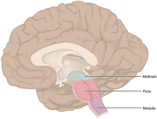
The name pons (bridge) stems from the fact that the pons provides a connection between the medulla oblongata, the cerebellum and the mesencephalon. Together, the pons and medulla oblongata border the rhomboid fossa (see below), the base of the fourth ventricle. The cerebellum is connected to the pons via the pedunculus cerebellaris medius.
The exit points of the VII (n. facialis) and VIII (n. vestibulocochlearis) cranial nerve are found at the location where the pons, medulla oblongata and the cerebellum meet. This point is also called the cerebellopontine angle. The VI cranial nerve (n. abducens) and V cranial nerve (n. trigeminus) also exit in the area of the pons.
The bridge nuclei (nuclei pontis) and several cranial nerve nuclei are located inside the pons. The cranial nerve nuclei located here are the abducens nucleus, the facial nucleus, the vestibulocochlearis nucleus, the nucleus principalis n. trigemini and the nucleus motorius n. trigemini.
The nucleus salivatorius, which transmits parasympathetic influences, is found in the area of the facial nucleus.
Structure of the Myencephalon
Medulla Oblongata
The medulla oblongata forms the lower part of the brain stem and has a caudal connection to the spinal cord. By definition, it extends from the pons to the end of C1, the first cervical nerve. Its cranial segment forms the base of the fourth ventricle (fossa rhomboidea, see above) with the pons.
Contents of the rhomboid fossa (fossa rhomboidea)
The colliculus facialis, formed by the inner abducens nerve, is found in the middle of the rhomboid fossa. Lateral to this, the protrusion of the vestibularis nuclei forms the colliculi facialis, the area vestibularis, and the protrusion of the nucleus cochlearis posterior forms the tuberculum acusticum.
Furthermore, the parasympathetic nucleus areas of the X (n. vagus) and XII (n. hypoglossus) cranial nerve protrude caudally into the rhomboid fossa as the trigonum nervi vagi and trigonum nervi hypoglossi, respectively.
In addition, the area postrema (vomiting center) – which is one of the circumventricular organs and part of the formatio reticularis (see below) – is located in the caudal area of the rhomboid fossa.
Topography of the medulla oblongata
The medulla oblongata is cranially connected with the cerebellum via the velum medullare inferius (lower cerebellar velum) and the pedunculi cerebellares inferius.
The pyramids, which contain the tractus corticospinalis (pyramidal tracts), border the medula oblongata on the ventral side, i.e., toward the frontal lobe. A majority of the fibers of the pyramidal tracts cross over to the contralateral site (approx. 80%) at the level of the medulla oblongata, causing this location to be referred to as the decussatio pyramidum (pyramidal decussation).
The remaining fibers cross over later. Motor phenomena and failures on the left side of the body are due to damage to the right hemisphere because of this.
The olivary bodies (olivae), which are connected to the cerebellum via the tractus olivocerebellaris, are located next to the pyramids. Information between the motoric centres and the cerebellum is passed from here through the nucleus olivaris inferior of the olivary bodies.
The XII cranial nerve (n. hypoglossus) exits between the two olivary bodies in the sulcus preolivaris. Other cranial nerves that exit in the area of the olivary bodies are the IX (n. glossopharyngeas), the X (n. vagus) and the XI cranial nerve (n. accesorius).
The tuberculum gracile and the tuberculum cuneatum, which contain the nucleus gracilis and the nucleus cuneatus respectively, are located dorsal to the medulla oblongata. The tuberculum gracile lies medial to the tuberculum cuneatum and is the synapse of the epicritical afferents of the trunk and lower half of the body.
The crossover of the afferents from the arm and throat area occurs in the tuberculum cuneatum. The lemniscus medialis travels from the tuberculi to the contralateral thalamus as a cohesive efferent. The thalamus acts as the third synapse and the third neuron to subsequently forward the information to the somatosensory cortex.
Circulatory and respiratory center
Another functional center located next to the area postrema in the vicinity of the medulla oblongata is the circulatory and respiratory center.
In addition, vegetative centers for regulating heartbeat, the metabolism, the width of blood vessels and individual reflexes, e.g., coughing, are located here. These centers are functionally ascribed to the formatio reticularis (see below).
Structure of the Formatio Reticularis
The formatio reticularis serves to regulate motoric and vegetative functions. To this end, it extends across the entire brain stem and contains multiple regulatory centers and nuclei. It also plays a crucial role in the formation of the circadian rhythm.
Nuclei of the formatio reticularis
The nuclei of the formatio reticularis are – contrary to the nuclei of the cranial nerves – diffuse in their differentiation. The nuclei can be categorised into a medial and lateral group by their localisation and into other groups by the transmitter they contain.
The nuclei of the medial group contain rather large neurons, whereas those of the lateral group mainly consist of small neurons. The larger neurons form ascending and descending pathways with their axons, while the axons of the small neurons usually do not extend through the brain stem.
Serotonin, noradrenaline, adrenaline, acetylcholine and dopamine are among the possible transmitters of the nuclei.
One example of a serotoninergic nucleus are the raphe nuclei, which extend into the hypothalamus, the neocortex and the limbic system. They are located both in the medulla oblongata and the pons as well as the mesencephalon, whereby they are each located on either side of the midline. They play a crucial role in the modulation of emotions and moods. A release of serotonin also results in deep sleep.
The locus caeruleus contains the transmitter noradrenaline and modulates the controls of motor processes in the cerebellum area. The release of noradrenaline mainly occurs while awake. It sends a stimulation throughout the entire cortex area of the brain, resulting in increased alertness.
The transmitter dopamine is mainly located in the area tegmentalis ventralis, which is found in the mesencephalon. The fibers chiefly project into the limbic system, which includes the amygdala and the hippocampus, and into the mesolimbic system (hypothalamus). Dopamine – transmitted through the nucleus accumbens – has a euphoric effect, among others.
Nuclei in the vicinity of the formatio reticularis that contain acetylcholine include the nucleus pedunculopontinus tegmentali and the nucleus dorsalis tegmentalis. The cholinergic neurons are activated in the waking state and in REM sleep, although they are inactive during deep sleep.
The fibers of the formatio reticularis, which are responsible for the waking state, are jointly referred to as the “ascending reticular activating system” (ARAS). Damage to or disruption of these fibers can lead to a state of coma.
However, other systems are also involved in the formation of the waking state, aside from the formatio reticularis. In all, the locus caeruleus (transmitted by the transmitter noradrenaline), the cholinergic fibers from the basal forebrain (e.g., nucleus basalis of Meynert), histaminergic fibres from the thalamus (nucleus centromedianus) and glutamatergic fibers from the thalamus (intralaminar nuclei) have a waking effect.
Substances that inhibit the aforementioned systems can cause sedation or a reduced state of alertness. For instance, benzodiazepine or barbiturates decrease the activity of the formatio reticularis. Alcohol also has a suppressive effect on the activity of the ARAS.
Respiratory center of the formatio reticularis
Two groups of neurons regulate respiration. One is the inspiratory neurons, and the other is the expiratory neurons.
The rhythmic alteration between inspiration and expiration is regulated by a cellular complex located in the vicinity of the ventral medulla oblongata (pneumotaxic center). This complex is called the pre-Bötzinger complex. Failure of this cellular complex results in breathing disorders.
Circulatory center of the formatio reticularis
The neurons for circulatory regulation are divided into blood pressure-increasing and -decreasing neurons. Multiple neurons with the same function are found in specific regions. The neurons that raise blood pressure tend to be laterally located.
The circulatory center is regulated by fibers from the hypothalamus, which pass through the fasciculus medialus telencephali (medial forebrain bundle).
Structure of the area postrema (vomiting center)
The area postrema is one of the circumventricular organs and is also located in the area of the formatio reticularis.
The fact that the blood-brain barrier is usually interrupted here, whereby substances from the blood can directly pass into the cerebral parenchyma (and vice versa), is characteristic of the circumventricular organs.
Certain substances can trigger a vomiting reaction, and so the stimulus is transmitted by a serotoninergic receptor of the sub-type 5-HT3. An increased concentration of serotonin in the blood, e.g., as a result of chemotherapy, can thus stimulate these receptors, thereby inducing a vomiting response. The vomiting stimulus can in turn be effectively suppressed by 5-HT3antagonists.
Two other receptors located in the area postrema, the stimulation of which results in illness and vomiting, are the dopaminergic D2 receptor and the histamine receptor of the type H1. Antagonists can thus also be applied as antiemetics against D2 and H1 receptors.
Gaze centers of the formatio reticularis
The gaze centers are divided into the pontine and vertical gaze center. The pontine gaze center, which regulates horizontal movements of the bulbus, is made of the paramedian pontine reticular formation (PPRF), whereas the vertical gaze center comprises the rostral interstitial nucleus of the medial longitudinal fasciculus (riFLM).
The medial longitudinal fasciculus connects the three eye muscle nuclei of cranial nerves III, IV and VI, and both sides run ventral to the aquaeductus mesencephali up to the cervical spinal cord.
Damage to the medial longitudinal fasciculus results in internuclear ophthalmoplegia (INO), the most common cause of which being damage to the bundle from multiple sclerosis.
A clinical symptom of internuclear ophthalmoplegia is that, when looking to the side, the contralateral eye remains in the center, and the ipsilateral eye exhibits monoocular nystagmus. The convergence reaction, however, is regular.
Review Questions
The solutions can be found beneath the bibliography.
1. Which of these statements about the rhombencephalon is not true?
- It develops from one of the cerebral vesicles.
- The rhombencephalon includes the cerebellum, pons and medulla oblongata.
- The cerebellum and pons are part of the metencephalon.
- The myelencephalon is formed by the medulla oblongata.
- Together, the mesencephalon and myencephalon form the rhombencephalon.
2. Which of these statements about the cerebellum is true?
- The cerebellum consists of the vestibulo-, spino- and pontocerebellum.
- The granular cells send their information through the transmitter GABA.
- From the inside out, the cerebellar cortex is made up of the stratum moleculare, the stratum granulosum and the stratum purkinjese.
- The pedunculus cerebellaris medius has two efferents.
- The ncl. dentatus, ncl. emboliformus, ncl. caudatus and ncl. globosus are the four cerebellar nuclei.
3. Which of these statements on the formatio reticularis is not true?
- The nuclei of the formatio reticularis are spread throughout it diffusely.
- Benzodiazepine leads to increased activity of the formatio reticularis.
- The raphe nuclei are serotoninergic.
- The locus caeruleus contains the transmitter noradrenaline.
- The ascending reticular activating system (ARAS) is part of the formatio reticularus.


Your blog is very nice. Wish to see much more like this.
ResponderEliminarhttps://blog.mindvalley.com/hindbrain
Thanks for this post. I have been looking for something like this for year’s thank you!
ResponderEliminarhttps://blog.mindvalley.com/hindbrain-function