Biology for Physicians: The Basics of Medical Microbiology
Table of Contents
Bacteria
All living organisms are divided into three domains:
- Eukaryotes (living organisms with a nucleus): humans, animals, plants, fungi
- Prokaryotes (living organisms without a nucleus): bacteria
- Archaea: formerly called “archaebacteria”
Protists combine the groups of unicellular pathogens with a nucleus (i.e. eukaryotes). Among the protists are:
- Protozoa (important pathogens: malaria, sleeping sickness)
- Unicellular algae
- Certain fungi
Morphology and structure of bacteria
Bacteria are prokaryotes: They do not have a enveloped nucleus. Instead, the DNA is compressed into a nucleus-like body, the nucleoid, without being limited by a membrane. There are also no other organelles like in eukaryotes (no ER, no mitochondria, etc.): the entire metabolism of the bacterium takes place in the cytoplasm.
The two most important characteristics that identify bacteria microscopically are the external appearance of the bacterium, the Gram stain, as well as its type of flagellation (if any).
The following table provides an overview of the morphological classification of bacteria.
Image: “Arrangements of cocci. (a) streptococcus, divide along same plane. (b) diplococcus, make pairs of two cocci after division. (c) tetrad, divide along two planes regularly. (d) sarcina, divide along three planes regularly. (e) staphylococcus, divide along planes irregularly.” by Y tambe. License: CC BY-SA 3.0
Bacterial cell walls
The bacterial cell wall attaches to the outside of the cell membrane. It provides mechanical stability and allows for the exchange of nutrients and waste. The peptidoglycan murein is part of almost all bacterial walls. Murein consists of N-acetyl glucosamine and N-acetylmuramic acid cross-linked through oligopeptides A murein sacculus develops from this linkage, which protects the cell and acts against osmotic pressure.
Classification of bacteria according to Gram staining
For the classification of bacteria, Gram staining, a method named after the Danish bacteriologist Hans Christian Gram (1853–1938), is used. It plays an important part in diagnostics because gram-negative bacteria are less sensitive to penicillin due to their additional lipid bilayer membrane.
Process of Gram staining:
- Administering the violet dye to the cells
- Washing off the stain with alcohol
- Red counter-staining of cells
Gram-positive bacteria: violet, Gram-negative bacteria: red
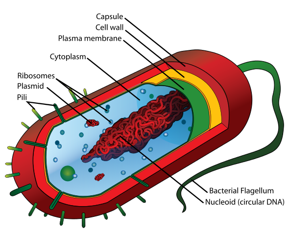
Image: “The cell envelope comprises a plasma membrane, seen here in green, and a thick peptidoglycan-containing cell wall (the yellow layer). No outer lipid membrane is present, as would be the case in Gram-negative bacteria. The red layer, known as the capsule, is distinct from the cell envelope.” License: CC0 1.0
Gram-positive bacteria have a thicker murein layer. In comparison, gram-negative bacteria have a thinner murein layer but instead a second membrane in the form of a lipid bi-layer.
For the hosts themselves, an infection with gramnegative bacteria is more critical. Upon decomposition, the lipopolysaccharides of the second membrane layer can be released by the host body as endotoxins. Endotoxins act as pyogenes and cause high fever and chills.
Overview of gram-positive and gram-negative bacteria and diseases
Bacterial flagella and pili
Approximately 50 % of prokaryotes move with the help of flagella. Flagella can either be monotrich, meaning one single flagellum, or polytrich, meaning more than one flagellum on the bacterium. The bacterial flagella consist of the protein flagellin and are not covered by the cell membrane. The placement of the flagella is either monopolar (at one end of the cell), bipolar (on both ends of the cell), or peritrich, (all over the bacterium).
Sometimes the proteins of the flagella are species-specific and thus work as antigenes, which can be identified in serology (i.e. in the case of salmonella or E. coli).
Pili are similar in structure to flagella but they are much smaller. Pili (or fimbria) help the bacterium better adhere to surfaces and other bacteria. This adherence of the bacterium to a potential host is made possible by the fimbria. Therefore, they represent a virulence factor.
Bacterial metabolism
Almost all bacteria need organic substances in order to survive and therefore belong to the heterotrophic organisms. Bacteria are further classified according to their relationship to oxygen.
Bacterial genetics
Bacteria are capable of exchanging genetic material with other bacteria. Due to the fact that bacteria can integrate foreign DNA into their own genome, they can re-combine their existing gene pool. This way, different bacteria strains can transfer genetic properties among each other.
The three mechanisms of gene transfer
- Conjugation: parasexual transfer through contact via F pili
- Transduction: gene transfer through bacteriophages (viruses that infect bacteria)
- Transformation: the introduction of free, isolated, foreign DNA into the bacterial genome
The cause of the pathogenic effect of bacteria
Different factors have an influence on the virulence (strength of pathogenicity) of bacteria.
- The number of infecting bacteria
- Adhesions
- Invasion factors
- Replication rate
- Formation of endo- and exotoxins
- The ability to avoid the immune system
Endotoxins: Endotoxins are created during the breakdown of parts of the bacterial cell wall (see above) when bacteria die. Through cytokines, endotoxins activate the complement cascade as well as the coagulation cascade in the host, which can result in septic shock. There are disease-unspecific symptoms caused by endotoxins such as: fever, pain, shock, fatigue, and discomfort.
Exotoxins: Some bacteria can produce toxins on their own and secrete them. If they target a host, this can result in very severe disease-specific symptoms. Examples are the cholera toxin, the botulinum toxin, the diphtheria toxin and the tetanus toxin.
Antibacterial substances: Antibiotics and chemotherapeuticals
Bactericidal substances kill bacteria, bacteriostatic substances simply inhibit their growth. Antibiotics are substances synthesized from bacteria and fungi, which work against other microorganisms. Antibiotics and chemotherapeuticals interfere in certain steps of the bacterial cell metabolism. They inhibit replication, transcription, and translation or damage the bacterial cell wall or cell membrane. Penicillins, for instance, inhibit the murein synthesis.
Fungi
Fungi belong to the eukaryote domain and, just like plants, have cells walls, vacuoles, exhibit cytoplasmic streaming, and are immobile. Almost all fungi, however, consist of chitin in their cell walls, not of cellulose. Fungi do not carry out photosynthesis but get their substrates for metabolism because they are saprophytes (obtain their food from dead matter).
Fungal growth forms and reproduction
Fungi are mostly composed of thread-like hyphae. The organism of a fungus consists of a tubular system in which the hyphae form a strongly branching net, the mycelium. Yeasts are the exception that do not form any hyphae nor a mycelium.
Fungi can reproduce sexually or asexually. Asexual reproduction takes place through binary fission, the breakdown of hyphae, budding (in yeasts), or the formation of conidia (containing asexual, mitotic spores). Sexual reproduction occurs through the merging of two physiologically different cells. The emerging diploid cell can now bud out and form diploid cells. If the reduction division does not occur until the zygote stage, haploid spores form.
Toxic synthesis products of fungi
Some fungi can produce toxic substances which pose a threat for humans. The following overview summarizes the most important fungi, the toxic substances they produce, as well as their effect.
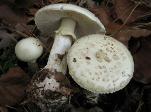
Image: “Knollenblätterpilz Heidelberg Deutschland” by Grossbildjaeger. License: CC BY-SA 3.0
The pathogenic effect of fungi
The infectious diseases caused by fungi are called mycoses. In a healthy individual, they usually do not pose a problem. If the immune system is compromised, however, they will break out as opportunistic infections.
- Dermatophytes: keratinophilic fungi that affect human skin, hair, and nails
- Yeast and mold fungi affect the gastrointestinal and respiratory mucous membranes (high risk after treatment with antibiotics!)
- Systemic mycoses develop when fungi spores are inhaled which can then manifest in different inner organs: this results in severe infections that can be lethal. (HIV-positive patients are suseptible to these infections.)
Synthesis of antibiotics
Flemming determined in 1928 that some fungi are capable of producing substances that are effective as antibiotics: penicillin from Penicillium notatum, cephalosporin from Acremonium and Griseofulvin from Penicillium griseofulvum. 50 of these approximately 2,000 substances characterized as antibiotics are used as chemotherapeuticals.
Viruses
Viruses are infectious particles that are between 20 nm and 300 nm in size. Viruses can neither grow on their own, nor can they reproduce. They use host cells instead. They invade them and use the host metabolism for their own reproduction. Bacteriophages are a particular type of viruses: They use bacteria as their hosts by injecting their genome into the bacterium and integrating it into the genome.
Structure and classification of viruses
The genetic information of the virus exists in the form of a nucleoid from DNA or RNA. The nucleoid is surrounded by capsid, a protein layer, and partially by an additional layer of lipids and glycoproteins. Some viruses have enzymes, for example, reverse transcriptase.
Viruses are classified according to the following characteristics:
- RNA or DNA?
- Single- or double-stranded genome?
- Naked or enveloped (additional envelope)?
- Cube-based, helical or complex capsid symmetry?
- Animals, plants, or humans as host?
- Immunological characteristics?
- Sensitive toward chemical or physical properties?
Viral replication
With regard to the type of replication, one has to differentiate between bacteriophages and eukaryotic viruses.
The replication cycle of bacteriophages
Phages are composed of a single- or double-stranded head and a tail, which serves to adhere to the host bacterium. After penetrating the cell wall, this hollow tail injects its genome into the bacterium. Afterwards, two different cycles can follow: The lytic replication cycle and the temperate/lysogenic cycle.
During the lytic replication cycle, DNA is transcribed immediately. The protein structures and the envelopes of the phage are replicated by the host, so is the DNA later on. The bacterium dies off in the process.
The temperate cycle is also referred to as lysogenic cycle. The term lysogenic describes the integration of the phage DNA into the bacteria chromosome. Prophages are created which are first replicated together with the bacteria DNA and inherited.
The replication cycle of viruses infecting eukaryotic cells
In this case, the viruses completely invade the host cell. The genome is released inside the infected cell.
The six steps of viral replication:
- Attachment: using specialized proteins to attach to the host cell
- Penetration: invading the cell
- Uncoating: the capsid is dismantled and genetic material made available
- Replication: the viral nucleic acid and viral proteins are synthesized
- Maturation: synthesis components form new viruses
- Liberation/Release: new viruses exit the cell via membrane lysis or pinching off the cell membrane
Special case: Retroviruses
In retroviruses, the genetic information is present in the form of RNA rather than DNA. Retroviruses possess a special enzyme, the reverse transcriptase (RT). The RT transcribes the RNA into DNA before releasing it into the host cell. The most prominent example for a retrovirus is HIV which causes AIDS.
Prions
The term prions is derived from the term proteinaceous infectious particle, which provides its own definition: Prions are very small, pathogenic infectious proteins. Prions are associated with several degenerative diseases, for example, bovine spongiform encephalopathy (BSE), commonly known as “mad cow disease”.
Other diseases include scrapie in sheep, Creutzfeld-Jakob disease and Kuru (found exclusively in a tribe practicing cannibalistic rites). The transfer mechanism of prions has not been determined as of yet.
Parasites
Parasites are organisms that take metabolic advantage of another organism. They can be classified as Viruses, Bacteria, Fungi, Protozoa, Helminths, or Arthropods.
Zoonoses- These are human diseases that are transmitted via animals.
Protozoa
Basic single-celled eukaryotes, consisting of genetic material and single-layer lipid membrane. They use arthropod vectors to infect species and can exist in two forms: an active trophozoite and a dormant cyst.
Classification
Helminths (worms)
Multi-celled organisms that can reproduce sexually or are hermaphroditic. They have the ability to develop into dormant cysts. Helminths live complex lifestyles involving animal and environmental reservoirs. They can be transmitted via fecal-oral, fecal-skin, or ingestion. Disease burden is directly related to the amount of worms that are in the host.
Nematodes (roundworms)
These are non-segmented worms that can infect the intestine, blood, or skin.
Flatworms (Platyhelminthes)
More primitive worm which are asymmetrical in shape. They are divided into trematodes (shistosoma) and cestodes (tapeworms).
Trematodes
A common water-borne parasite. It is estimated that between 200—300 million people worldwide infested with this parasite. They are laid as eggs in fresh water. These parasites cannot swim and need to mature in freshwater snails. Once matured, the shistosoma penetrate through exposed human skin into the bloodstream. The trematodes will enter the venous system and lay eggs into the intestinal tract. The eggs are released with the human feces into the environment.
Cestodes
Segmented worm that lives in the digestive tract of its host and absorbs nutrients as they pass through the intestine. The parasite eggs are ingested through undercooked food.
Prevalence of parasitic infections
Majority of cases occur in the tropical region and in under-developed countries. Hosts might not show any symptoms of infestation until host becomes immunosuppressed.
Review Questions
The correct answers can be found below the references.
1. Over the course of an infection disease, an endotoxin shock can occur as a result of the release of lipopolysaccharides. During which infection is this most likely?
- Retroviruses
- Gram-negative rods
- Gram-positive rods
- Gram-positive diplococci
- Facultative anaerobes
2. A 13-year old boy who has never been seriously ill before is taken to the hospital with a fever, difficulty breathing, and increasing suspicion of pneumonia. Four days prior, his family physician had prescribed a broad-spectrum antibiotic (penicillin with gram-negative and gram-positive efficacy spectrum) without any improvement. Therapy failure as well as the clinical symptoms the boy is displaying point to which most likely original pathogen?
- Staphylococci
- Enterobacteriaceae
- Viruses
- Mycoplasmas
- Streptococci
3. Retroviridae represent a certain group of viruses. Which stage of the replication cycle of viruses was the determining factor for the name of these special viruses?
- Penetration
- Transcription
- Maturation
- Uncoating
- Liberation/Release


Comentários
Enviar um comentário