Subclavian Arteries
Table of Contents
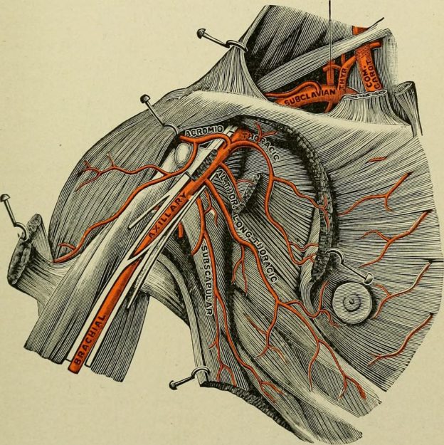
Image: “Subclavian and axillary arteries” by Internet Archive Book Images – https://www.flickr.com/photos/internetarchivebookimages/14764575614/ Source book page: https://archive.org/stream/textbookofanatom00bund/textbookofanatom00bund#page/n184/mode/1up, License: Flickr API
Origin of the Subclavian Arteries
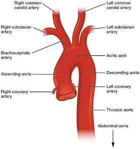
The right and left subclavian arteries have different origins:
Right subclavian artery (RSA)
The right brachiocephalic artery divides into two terminal branches: right subclavian artery and right common carotid artery.
Left subclavian artery (LSA)
The arch of aorta gives off three branches: brachiocephalic artery, left common carotid artery and left subclavian artery. Left subclavian is the last branch of the arch of aorta.
Course of the Subclavian Arteries
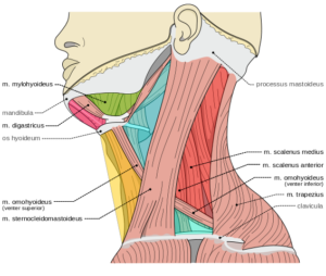
The subclavian artery exits the thorax and enters the neck by passing through the superior thoracic aperture (thoracic outlet). Superior thoracic aperture lies between the anterior and middle scalene muscles. It passes anterior to the first rib and underneath the clavicle. It is named “axillary artery” just after passing the lateral border of the first rib.
Parts of the Subclavian Artery
On basis of its relation with the anterior scalene muscle, the subclavian artery is divided into three parts.
First part of the artery starts at its origin up until the medial margin of the scalenus muscle. Second part starts from the medial margin of the scalenus muscle and ends at the lateral margin. Third part continues from the lateral margin of the scalenus muscle and ends at the outer border of the first rib. Relations of all these parts of the artery are mentioned below.
First part of the right subclavian artery
The first part of the right subclavian artery originates from the brachiocephalic trunk, just posterior to the upper part of the right sternoclavicular joint. It moves in upward and lateral direction towards the medial border of the scalenus anterior muscle. Its course varies from person to person.
Anterior relations of the first part of the artery include skin, superficial fascia, platysma muscle, deep cervical fascia, the sternocleidomastoid muscle, the sternohyoid muscle and sternothyroid muscle.
This part of the artery is also crossed by the internal jugular and vertebral veins and the vagus nerve accompanies the internal jugular vein. Cardiac branches of the vagus nerve also cross this part. The sympathetic trunk forms a ring around the first part of the vessel called the subclavian loop. The anterior jugular vein (also called external jugular vein), which courses superficially in the center of the neck, is separated from the artery by the sternohyoid and sternothyroid muscles.
Posterior relations include the thoracic pleura at the apex of the lungs, the sympathetic trunk, the longus collis muscle and the first thoracic vertebra. The right recurrent laryngeal nervewinds around the lower and posterior part of the artery.
First part of the left subclavian artery
On the left side, the subclavian artery has a slightly different origin and course. The first part of the left subclavian artery starts from the arch of the aorta. The artery moves upwards into the superior mediastinal cavity reaching the root of the neck. There, it arches toward the lateral side reaching the medial border of the scalenus anterior muscle.
The Vagus nerve along with its cardiac branches and the phrenic nerve are the anterior relations of this artery. The left common carotid artery, left internal jugular and vertebral veinstravel parallel to the artery. Left subclavian artery is covered by the sternothyroid, sternohyoid, and sternocleidomastoid muscles.
Posterior relations include the esophagus, thoracic duct, left recurrent laryngeal nerve, inferior cervical ganglion of the sympathetic trunk and Longus colli muscle.
Moving upwards, it comes into relationship with the esophagus and the thoracic duct. The thoracic duct makes an arch over the artery and drains the lymph into the blood circulation at the junction of the subclavian and the internal jugular veins. Medial relations of the artery include the esophagus, trachea, thoracic duct and left recurrent laryngeal nerve, whereas the lateral relations include the left pleura and lung.
Second part of the subclavian artery
The second part of the subclavian artery lies behind the scalenus anterior. It is relatively shorter in length.
Anterior relations include the skin, superficial fascia, platysma, deep cervical fascia, sternocleidomastoid and scalenus anterior. The Phrenic nerve is also close to this part of the artery, as it is separated by the scalenus anterior on the right side of the neck, while on left side it crosses the first part of the artery near the medial border of the muscle.
Posterior relations include the pleura and the scalenus medius. Superiorly lies the brachial plexus of nerves and inferiorly lies the pleura. The subclavian vein, separated by the scalenus anterior muscle, lies below the artery.
Third part of the subclavian artery
The third part of the artery starts from the lateral border of the scalenus anterior muscle up until the outer border of the first rib, here onwards it continues as the axillary artery. The third part of the subclavian artery is the most superficial part of it. It lies in the subclavian triangle.
It is covered by the skin, the superficial fascia, the platysma, the supraclavicular nerves, and the deep cervical fascia. It is crossed by the external jugular vein, which receives the transverse scapular, transverse cervical and anterior jugular veins during its course; this leads to the formation of a venous plexus anterior to the artery.
The nerve to the subclavius muscle moves downwards anterior to the artery, just posterior to the veins. Behind the clavicle and the subclavius muscle, the subclavian artery terminates and becomes the axillary artery. It is crossed by the transverse scapular vessels before termination.
The subclavian vein lies anteriorly and slightly inferior to the artery. Posterior to the artery is the lowest trunk of the brachial plexus. Superolaterally lie the upper trunks of the brachial plexus and the omohyoideus muscle. Inferior relations include the upper surface of the first rib, on which the artery rests.
Branches of the Subclavian Artery
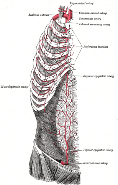
Image: “Internal thoracic (mammary) artery” by Henry Vandyke Carter, Henry Gray (1918) in “Anatomy of the Human Body”, Bartleby.com: Gray’s Anatomy, Plate 522, License: Public Domain
The branches of the artery are described one by one from each part of the artery:
- From the first part
- Vertebral artery: This branch of the first part of the subclavian artery moves superiorly towards the base of the skull and becomes part of the circle of Willis. It supplies blood to the posterior cerebral circulation.
- Internal thoracic artery: This branch of the first part moves inferiorly into the chest and supplies various parts of the chest wall and mediastinum.
- Thyrocervical trunk: This further divides into many branches soon after it emerges from the subclavain artery.
- From the second part
- Costocervical trunk courses cranially before bifurcating.
- From the third part
- Dorsal scapular artery courses posteriorly.
Variant Anatomy
Aberrant right subclavian artery is the most common variant of the subclavian artery. This vessel originates distally to the left subclavian artery. It moves posteriorly between the trachea and esophagus. It can lead to compression of the trachea or esophagus making breathing difficult or difficulty in swallowing (dysphagia lusoria, or ‘trouble swallowing because of a trick of nature’).
An aberrant left subclavian artery can also originate from the right side of the aortic arch.


Comentários
Enviar um comentário